{"title":"默认模式网络的活动介导了外周浆胶质细胞系源性神经营养因子水平对重度抑郁症患者反刍的影响。","authors":"Fennan Jia, Xiao Chen, Xingran Wang, Chuansheng Quan, Jing Ruan, Yuexiang Huang, Xiaoqian Fu, Yan Wang, Hongyan Sun, Lili Liu, Yuan Zhou, Chaogan Yan, Yansong Liu, Xiangdong Du","doi":"10.1093/psyrad/kkaf014","DOIUrl":null,"url":null,"abstract":"<p><strong>Background: </strong>Rumination is a pivotal psychopathological process in major depressive disorder (MDD). The neurotrophic hypothesis suggests that glial cell line-derived neurotrophic factor (GDNF) might play a role in brain dysfunction and clinical symptoms of MDD. However, the relationship remains unclear.</p><p><strong>Methods: </strong>Thirty-three individuals with MDD and 33 healthy controls (HCs) underwent functional magnetic resonance imaging (fMRI) while performing a rumination state task designed to induce sustained, active rumination. The Ruminative Response Scale (RRS) was administered to assess individual rumination tendency. Brain activity within the default mode network (DMN) subsystems during rumination was characterized using both fractional amplitude of low-frequency fluctuations (fALFF) and functional connectivity (FC) analyses. Serum levels of GDNF and inflammatory markers [interleukin (IL)-6, IL-8, and C-reactive protein] were quantified in all participants. We then examined the relationships between regional brain activity (fALFF values), GDNF levels, and rumination severity (RRS scores) in the MDD group.</p><p><strong>Results: </strong>Compared to HCs, MDD patients exhibited significantly reduced serum levels of both GDNF (<i>t = -</i>3.204, <i>P</i> = 0.002) and IL-8 (<i>t</i> = -3.239, <i>P </i>= 0.002). Significant interaction effects were observed in fALFF within both the dorsal medial prefrontal cortex (DMPFC; <i>F </i>= 25.075, <i>P < </i>0.001) and medial temporal lobe (MTL; <i>F </i>= 28.753, <i>P </i>< 0.001) subsystems of the DMN. Mediation analysis revealed that the relationship between GDNF levels and brooding rumination in MDD patients was mediated by neural activity within the DMPFC subsystem.</p><p><strong>Conclusions: </strong>In MDD patients, GDNF levels were associated with neural activity within the DMPFC subsystem of the DMN, which statistically mediated the link to rumination severity.</p>","PeriodicalId":93496,"journal":{"name":"Psychoradiology","volume":"5 ","pages":"kkaf014"},"PeriodicalIF":2.9000,"publicationDate":"2025-05-28","publicationTypes":"Journal Article","fieldsOfStudy":null,"isOpenAccess":false,"openAccessPdf":"https://www.ncbi.nlm.nih.gov/pmc/articles/PMC12202882/pdf/","citationCount":"0","resultStr":"{\"title\":\"Activity of the default mode network mediates the effect of peripheral plasma glial cell line-derived neurotrophic factor levels on rumination in major depressive disorder patients.\",\"authors\":\"Fennan Jia, Xiao Chen, Xingran Wang, Chuansheng Quan, Jing Ruan, Yuexiang Huang, Xiaoqian Fu, Yan Wang, Hongyan Sun, Lili Liu, Yuan Zhou, Chaogan Yan, Yansong Liu, Xiangdong Du\",\"doi\":\"10.1093/psyrad/kkaf014\",\"DOIUrl\":null,\"url\":null,\"abstract\":\"<p><strong>Background: </strong>Rumination is a pivotal psychopathological process in major depressive disorder (MDD). The neurotrophic hypothesis suggests that glial cell line-derived neurotrophic factor (GDNF) might play a role in brain dysfunction and clinical symptoms of MDD. However, the relationship remains unclear.</p><p><strong>Methods: </strong>Thirty-three individuals with MDD and 33 healthy controls (HCs) underwent functional magnetic resonance imaging (fMRI) while performing a rumination state task designed to induce sustained, active rumination. The Ruminative Response Scale (RRS) was administered to assess individual rumination tendency. Brain activity within the default mode network (DMN) subsystems during rumination was characterized using both fractional amplitude of low-frequency fluctuations (fALFF) and functional connectivity (FC) analyses. Serum levels of GDNF and inflammatory markers [interleukin (IL)-6, IL-8, and C-reactive protein] were quantified in all participants. We then examined the relationships between regional brain activity (fALFF values), GDNF levels, and rumination severity (RRS scores) in the MDD group.</p><p><strong>Results: </strong>Compared to HCs, MDD patients exhibited significantly reduced serum levels of both GDNF (<i>t = -</i>3.204, <i>P</i> = 0.002) and IL-8 (<i>t</i> = -3.239, <i>P </i>= 0.002). Significant interaction effects were observed in fALFF within both the dorsal medial prefrontal cortex (DMPFC; <i>F </i>= 25.075, <i>P < </i>0.001) and medial temporal lobe (MTL; <i>F </i>= 28.753, <i>P </i>< 0.001) subsystems of the DMN. Mediation analysis revealed that the relationship between GDNF levels and brooding rumination in MDD patients was mediated by neural activity within the DMPFC subsystem.</p><p><strong>Conclusions: </strong>In MDD patients, GDNF levels were associated with neural activity within the DMPFC subsystem of the DMN, which statistically mediated the link to rumination severity.</p>\",\"PeriodicalId\":93496,\"journal\":{\"name\":\"Psychoradiology\",\"volume\":\"5 \",\"pages\":\"kkaf014\"},\"PeriodicalIF\":2.9000,\"publicationDate\":\"2025-05-28\",\"publicationTypes\":\"Journal Article\",\"fieldsOfStudy\":null,\"isOpenAccess\":false,\"openAccessPdf\":\"https://www.ncbi.nlm.nih.gov/pmc/articles/PMC12202882/pdf/\",\"citationCount\":\"0\",\"resultStr\":null,\"platform\":\"Semanticscholar\",\"paperid\":null,\"PeriodicalName\":\"Psychoradiology\",\"FirstCategoryId\":\"1085\",\"ListUrlMain\":\"https://doi.org/10.1093/psyrad/kkaf014\",\"RegionNum\":0,\"RegionCategory\":null,\"ArticlePicture\":[],\"TitleCN\":null,\"AbstractTextCN\":null,\"PMCID\":null,\"EPubDate\":\"2025/1/1 0:00:00\",\"PubModel\":\"eCollection\",\"JCR\":\"\",\"JCRName\":\"\",\"Score\":null,\"Total\":0}","platform":"Semanticscholar","paperid":null,"PeriodicalName":"Psychoradiology","FirstCategoryId":"1085","ListUrlMain":"https://doi.org/10.1093/psyrad/kkaf014","RegionNum":0,"RegionCategory":null,"ArticlePicture":[],"TitleCN":null,"AbstractTextCN":null,"PMCID":null,"EPubDate":"2025/1/1 0:00:00","PubModel":"eCollection","JCR":"","JCRName":"","Score":null,"Total":0}
引用次数: 0
摘要
背景:反刍是重度抑郁障碍(MDD)的关键精神病理过程。神经营养假说提示神经胶质细胞系来源的神经营养因子(GDNF)可能在重度抑郁症的脑功能障碍和临床症状中起作用。然而,这种关系尚不清楚。方法:33名重度抑郁症患者和33名健康对照(hc)在执行反刍状态任务时进行功能磁共振成像(fMRI),以诱导持续、主动的反刍。采用反刍反应量表(RRS)评估个体反刍倾向。利用低频波动分数幅值(fALFF)和功能连通性(FC)分析,对反刍过程中默认模式网络(DMN)子系统中的大脑活动进行了表征。对所有参与者的血清GDNF和炎症标志物[白细胞介素(IL)-6、IL-8和c反应蛋白]水平进行量化。然后,我们检查了MDD组的区域脑活动(fALFF值)、GDNF水平和反刍严重程度(RRS评分)之间的关系。结果:与hcc患者相比,MDD患者血清GDNF (t = -3.204, P = 0.002)和IL-8 (t = -3.239, P = 0.002)水平均显著降低。在背内侧前额叶皮层(DMPFC;F = 25.075, P 0.001)和内侧颞叶(MTL;结论:在MDD患者中,GDNF水平与DMN DMPFC子系统内的神经活动相关,这在统计学上介导了反刍严重程度的联系。
Activity of the default mode network mediates the effect of peripheral plasma glial cell line-derived neurotrophic factor levels on rumination in major depressive disorder patients.
Background: Rumination is a pivotal psychopathological process in major depressive disorder (MDD). The neurotrophic hypothesis suggests that glial cell line-derived neurotrophic factor (GDNF) might play a role in brain dysfunction and clinical symptoms of MDD. However, the relationship remains unclear.
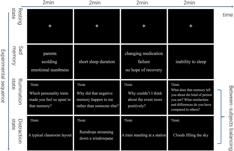
Methods: Thirty-three individuals with MDD and 33 healthy controls (HCs) underwent functional magnetic resonance imaging (fMRI) while performing a rumination state task designed to induce sustained, active rumination. The Ruminative Response Scale (RRS) was administered to assess individual rumination tendency. Brain activity within the default mode network (DMN) subsystems during rumination was characterized using both fractional amplitude of low-frequency fluctuations (fALFF) and functional connectivity (FC) analyses. Serum levels of GDNF and inflammatory markers [interleukin (IL)-6, IL-8, and C-reactive protein] were quantified in all participants. We then examined the relationships between regional brain activity (fALFF values), GDNF levels, and rumination severity (RRS scores) in the MDD group.
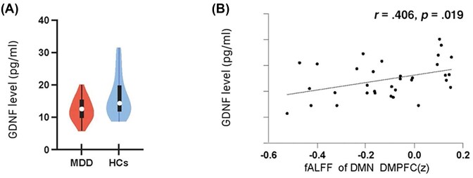
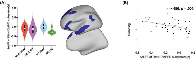
Results: Compared to HCs, MDD patients exhibited significantly reduced serum levels of both GDNF (t = -3.204, P = 0.002) and IL-8 (t = -3.239, P = 0.002). Significant interaction effects were observed in fALFF within both the dorsal medial prefrontal cortex (DMPFC; F = 25.075, P < 0.001) and medial temporal lobe (MTL; F = 28.753, P < 0.001) subsystems of the DMN. Mediation analysis revealed that the relationship between GDNF levels and brooding rumination in MDD patients was mediated by neural activity within the DMPFC subsystem.
Conclusions: In MDD patients, GDNF levels were associated with neural activity within the DMPFC subsystem of the DMN, which statistically mediated the link to rumination severity.

 求助内容:
求助内容: 应助结果提醒方式:


