Roland-Richard Macharzina, Simon Stemmler, Werner Vach, Thomas Winker, Jana Taron, Christopher L Schlett, Michael Weinbeck, Matthias Siepe, Martin Czerny, Fabian Bamberg, Thomas Zeller, Dirk Westermann, Martin Soschynski
{"title":"颈动脉斑块的超高分辨率和双能量计算机断层扫描通过新颖的体积分析来区分有症状和无症状的患者。","authors":"Roland-Richard Macharzina, Simon Stemmler, Werner Vach, Thomas Winker, Jana Taron, Christopher L Schlett, Michael Weinbeck, Matthias Siepe, Martin Czerny, Fabian Bamberg, Thomas Zeller, Dirk Westermann, Martin Soschynski","doi":"10.1093/icvts/ivaf158","DOIUrl":null,"url":null,"abstract":"<p><strong>Objectives: </strong>The indication for carotid endarterectomy (CEA) mainly relies on the degree of stenosis and neurological symptoms. Plaque vulnerability has been associated with stroke risk, but identification on single-energy computed tomography (CT) has yielded heterogeneous results and is not routinely applied to clinical diagnostics. Hence, we intended to analyse CEA specimens for vulnerability features using dual-source CT and correlate these features with the presence of preprocedural symptoms.</p><p><strong>Methods: </strong>CT was performed on 187 carotid plaque specimens using ultra-high-resolution and dual-energy imaging on a dual-source scanner. Plaques were separated into calcified versus non-calcified volumes and analysed concerning HU-density, calcifications and volumetric dual-energy indices (DEIs). Comparative statistical analysis of plaque characteristics was performed with respect to the presence of neurological symptoms.</p><p><strong>Results: </strong>The degree of stenosis of symptomatic and asymptomatic plaques was indifferent (69.2 ± 12.3% vs 66.3 ± 13.7%). The highest diagnostic accuracies were obtained by the % calcified volume (AUC 0.63 (0.54-0.71)), average whole plaque HU (AUC 0.71 (0.64-0.79)), profound calcification (AUC 0.74 (0.66-0.81)), calcification spots <1 mm (AUC 0.71 (0.63-0.79)) and spotty calcification (AUC 0.74 (0.66-0.82)). The diagnostic accuracy for symptomatic plaques was insignificant using average non-calcified plaque HU (AUC 0.59 (0.48-0.65)), but significant using average non-calcified plaque DEI (AUC 0.66 (0.58-0.74)).</p><p><strong>Conclusions: </strong>Symptomatic plaques were identified best by measuring density of the whole, calcified or non-calcified plaque and via spotty, profoundly localized and less dense calcification. A volumetric DEI identifies symptomatic plaques with non-calcified plaque characteristics more accurately than single-energy CT. Future clinical studies are necessary to confirm these findings in patients.</p>","PeriodicalId":73406,"journal":{"name":"Interdisciplinary cardiovascular and thoracic surgery","volume":" ","pages":""},"PeriodicalIF":0.0000,"publicationDate":"2025-07-03","publicationTypes":"Journal Article","fieldsOfStudy":null,"isOpenAccess":false,"openAccessPdf":"https://www.ncbi.nlm.nih.gov/pmc/articles/PMC12270255/pdf/","citationCount":"0","resultStr":"{\"title\":\"Ultra-high-resolution and dual-energy computed tomography of carotid artery plaques differentiate symptomatic and asymptomatic patients by novel volumetric analysis.\",\"authors\":\"Roland-Richard Macharzina, Simon Stemmler, Werner Vach, Thomas Winker, Jana Taron, Christopher L Schlett, Michael Weinbeck, Matthias Siepe, Martin Czerny, Fabian Bamberg, Thomas Zeller, Dirk Westermann, Martin Soschynski\",\"doi\":\"10.1093/icvts/ivaf158\",\"DOIUrl\":null,\"url\":null,\"abstract\":\"<p><strong>Objectives: </strong>The indication for carotid endarterectomy (CEA) mainly relies on the degree of stenosis and neurological symptoms. Plaque vulnerability has been associated with stroke risk, but identification on single-energy computed tomography (CT) has yielded heterogeneous results and is not routinely applied to clinical diagnostics. Hence, we intended to analyse CEA specimens for vulnerability features using dual-source CT and correlate these features with the presence of preprocedural symptoms.</p><p><strong>Methods: </strong>CT was performed on 187 carotid plaque specimens using ultra-high-resolution and dual-energy imaging on a dual-source scanner. Plaques were separated into calcified versus non-calcified volumes and analysed concerning HU-density, calcifications and volumetric dual-energy indices (DEIs). Comparative statistical analysis of plaque characteristics was performed with respect to the presence of neurological symptoms.</p><p><strong>Results: </strong>The degree of stenosis of symptomatic and asymptomatic plaques was indifferent (69.2 ± 12.3% vs 66.3 ± 13.7%). The highest diagnostic accuracies were obtained by the % calcified volume (AUC 0.63 (0.54-0.71)), average whole plaque HU (AUC 0.71 (0.64-0.79)), profound calcification (AUC 0.74 (0.66-0.81)), calcification spots <1 mm (AUC 0.71 (0.63-0.79)) and spotty calcification (AUC 0.74 (0.66-0.82)). The diagnostic accuracy for symptomatic plaques was insignificant using average non-calcified plaque HU (AUC 0.59 (0.48-0.65)), but significant using average non-calcified plaque DEI (AUC 0.66 (0.58-0.74)).</p><p><strong>Conclusions: </strong>Symptomatic plaques were identified best by measuring density of the whole, calcified or non-calcified plaque and via spotty, profoundly localized and less dense calcification. A volumetric DEI identifies symptomatic plaques with non-calcified plaque characteristics more accurately than single-energy CT. Future clinical studies are necessary to confirm these findings in patients.</p>\",\"PeriodicalId\":73406,\"journal\":{\"name\":\"Interdisciplinary cardiovascular and thoracic surgery\",\"volume\":\" \",\"pages\":\"\"},\"PeriodicalIF\":0.0000,\"publicationDate\":\"2025-07-03\",\"publicationTypes\":\"Journal Article\",\"fieldsOfStudy\":null,\"isOpenAccess\":false,\"openAccessPdf\":\"https://www.ncbi.nlm.nih.gov/pmc/articles/PMC12270255/pdf/\",\"citationCount\":\"0\",\"resultStr\":null,\"platform\":\"Semanticscholar\",\"paperid\":null,\"PeriodicalName\":\"Interdisciplinary cardiovascular and thoracic surgery\",\"FirstCategoryId\":\"1085\",\"ListUrlMain\":\"https://doi.org/10.1093/icvts/ivaf158\",\"RegionNum\":0,\"RegionCategory\":null,\"ArticlePicture\":[],\"TitleCN\":null,\"AbstractTextCN\":null,\"PMCID\":null,\"EPubDate\":\"\",\"PubModel\":\"\",\"JCR\":\"0\",\"JCRName\":\"CARDIAC & CARDIOVASCULAR SYSTEMS\",\"Score\":null,\"Total\":0}","platform":"Semanticscholar","paperid":null,"PeriodicalName":"Interdisciplinary cardiovascular and thoracic surgery","FirstCategoryId":"1085","ListUrlMain":"https://doi.org/10.1093/icvts/ivaf158","RegionNum":0,"RegionCategory":null,"ArticlePicture":[],"TitleCN":null,"AbstractTextCN":null,"PMCID":null,"EPubDate":"","PubModel":"","JCR":"0","JCRName":"CARDIAC & CARDIOVASCULAR SYSTEMS","Score":null,"Total":0}
引用次数: 0
摘要
目的:颈动脉内膜切除术(CEA)的适应症主要取决于狭窄程度和神经系统症状。斑块易感性与卒中风险相关,但单能量CT的识别结果不一致,不能常规应用于临床诊断。因此,我们打算使用双源计算机断层扫描(CT)分析CEA标本的易损性特征,并将这些特征与手术前症状的存在联系起来。方法:采用双源扫描仪对187例颈动脉斑块标本进行超高分辨率双能成像。将斑块分为钙化和非钙化体积,并分析其hu密度、钙化和体积双能量指数(DEI)。对斑块特征与神经症状的存在进行比较统计分析。结果:有症状斑块和无症状斑块狭窄程度无差异(69.2±12.3% vs 66.3±13.7%)。钙化体积% (AUC 0.63(0.54-0.71))、全斑块平均HU (AUC 0.71(0.64-0.79))、深度钙化(AUC 0.74(0.66-0.81))、钙化斑点的诊断准确率最高。结论:通过测量全斑块、钙化斑块或非钙化斑块的密度,以及通过点状、深度局部和低密度钙化来识别症状性斑块。容积DEI比单能量CT更准确地识别具有非钙化斑块特征的症状性斑块。需要进一步的临床研究来证实这些发现。
Ultra-high-resolution and dual-energy computed tomography of carotid artery plaques differentiate symptomatic and asymptomatic patients by novel volumetric analysis.
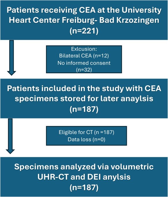
Objectives: The indication for carotid endarterectomy (CEA) mainly relies on the degree of stenosis and neurological symptoms. Plaque vulnerability has been associated with stroke risk, but identification on single-energy computed tomography (CT) has yielded heterogeneous results and is not routinely applied to clinical diagnostics. Hence, we intended to analyse CEA specimens for vulnerability features using dual-source CT and correlate these features with the presence of preprocedural symptoms.
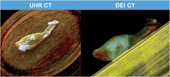
Methods: CT was performed on 187 carotid plaque specimens using ultra-high-resolution and dual-energy imaging on a dual-source scanner. Plaques were separated into calcified versus non-calcified volumes and analysed concerning HU-density, calcifications and volumetric dual-energy indices (DEIs). Comparative statistical analysis of plaque characteristics was performed with respect to the presence of neurological symptoms.
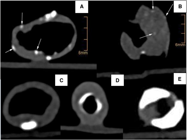
Results: The degree of stenosis of symptomatic and asymptomatic plaques was indifferent (69.2 ± 12.3% vs 66.3 ± 13.7%). The highest diagnostic accuracies were obtained by the % calcified volume (AUC 0.63 (0.54-0.71)), average whole plaque HU (AUC 0.71 (0.64-0.79)), profound calcification (AUC 0.74 (0.66-0.81)), calcification spots <1 mm (AUC 0.71 (0.63-0.79)) and spotty calcification (AUC 0.74 (0.66-0.82)). The diagnostic accuracy for symptomatic plaques was insignificant using average non-calcified plaque HU (AUC 0.59 (0.48-0.65)), but significant using average non-calcified plaque DEI (AUC 0.66 (0.58-0.74)).
Conclusions: Symptomatic plaques were identified best by measuring density of the whole, calcified or non-calcified plaque and via spotty, profoundly localized and less dense calcification. A volumetric DEI identifies symptomatic plaques with non-calcified plaque characteristics more accurately than single-energy CT. Future clinical studies are necessary to confirm these findings in patients.

 求助内容:
求助内容: 应助结果提醒方式:


