Sohee Park, Seung-Ah Lee, Jong Eun Lee, Joon-Won Kang, Dong Hyun Yang, Hyun Jung Koo
{"title":"利用ct应变参数分析主动脉狭窄严重程度对左心功能的影响。","authors":"Sohee Park, Seung-Ah Lee, Jong Eun Lee, Joon-Won Kang, Dong Hyun Yang, Hyun Jung Koo","doi":"10.3348/kjr.2024.1261","DOIUrl":null,"url":null,"abstract":"<p><strong>Objective: </strong>This study aimed to evaluate changes in left ventricular and left atrial mechanics in relation to the severity of aortic stenosis (AS) by comparing computed tomography (CT)-derived strain values in patients with mild-to-severe AS.</p><p><strong>Materials and methods: </strong>This single-center retrospective study included 120 patients (median age, 76 years; 45.0% male), comprising 30, 30, and 60 patients with mild, moderate, and severe AS, respectively, all of whom underwent multiphase cardiac CT between 2015 and 2021. Patients were selected from 177 individuals who met the initial eligibility criteria, with matching for age, sex, and hypertension in a 1:1:2 ratio across the mild, moderate, and severe AS groups. Electrocardiography-gated cardiac CT images were analyzed to obtain various quantitative left ventricle (LV) and left atrium (LA) strain parameters. Statistical differences in cardiac CT-derived LV and LA strain parameters among mild, moderate, and severe AS were evaluated using the Kruskal-Wallis test, followed by post-hoc tests.</p><p><strong>Results: </strong>The median LV global longitudinal strain differed significantly across AS severity (GLS: -19.4%, -18.2%, and -16.2% for mild, moderate, and severe AS, respectively; <i>P</i> < 0.001), with the absolute value decreasing as AS severity increased. Additionally, the median values of LV global circumferential strain (GCS: -29.8%, -30.8%, and -27.4%, respectively; <i>P</i> = 0.045), LV global radial strain (GRS: 50.1%, 50.3%, and 39.3%, respectively; <i>P</i> = 0.004), and LA conduit strain (11.5%, 11.2%, and 9.0%, respectively; <i>P</i> = 0.031) differed significantly according to AS severity, with lower absolute values observed in patients with severe AS.</p><p><strong>Conclusion: </strong>In patients with AS, CT-derived LV and LA strains revealed changes in myocardial deformation according to AS severity. Specifically, there was a gradual decrease in the absolute value of LV GLS with increasing AS severity and initial preservation until moderate AS, followed by an eventual decrease in the absolute values of LV GCS, LV GRS, and LA conduit strain in severe AS.</p>","PeriodicalId":17881,"journal":{"name":"Korean Journal of Radiology","volume":"26 7","pages":"626-637"},"PeriodicalIF":5.3000,"publicationDate":"2025-07-01","publicationTypes":"Journal Article","fieldsOfStudy":null,"isOpenAccess":false,"openAccessPdf":"https://www.ncbi.nlm.nih.gov/pmc/articles/PMC12235543/pdf/","citationCount":"0","resultStr":"{\"title\":\"Impact of Aortic Stenosis Severity on Left Heart Function Analyzed Using CT-Derived Strain Parameters.\",\"authors\":\"Sohee Park, Seung-Ah Lee, Jong Eun Lee, Joon-Won Kang, Dong Hyun Yang, Hyun Jung Koo\",\"doi\":\"10.3348/kjr.2024.1261\",\"DOIUrl\":null,\"url\":null,\"abstract\":\"<p><strong>Objective: </strong>This study aimed to evaluate changes in left ventricular and left atrial mechanics in relation to the severity of aortic stenosis (AS) by comparing computed tomography (CT)-derived strain values in patients with mild-to-severe AS.</p><p><strong>Materials and methods: </strong>This single-center retrospective study included 120 patients (median age, 76 years; 45.0% male), comprising 30, 30, and 60 patients with mild, moderate, and severe AS, respectively, all of whom underwent multiphase cardiac CT between 2015 and 2021. Patients were selected from 177 individuals who met the initial eligibility criteria, with matching for age, sex, and hypertension in a 1:1:2 ratio across the mild, moderate, and severe AS groups. Electrocardiography-gated cardiac CT images were analyzed to obtain various quantitative left ventricle (LV) and left atrium (LA) strain parameters. Statistical differences in cardiac CT-derived LV and LA strain parameters among mild, moderate, and severe AS were evaluated using the Kruskal-Wallis test, followed by post-hoc tests.</p><p><strong>Results: </strong>The median LV global longitudinal strain differed significantly across AS severity (GLS: -19.4%, -18.2%, and -16.2% for mild, moderate, and severe AS, respectively; <i>P</i> < 0.001), with the absolute value decreasing as AS severity increased. Additionally, the median values of LV global circumferential strain (GCS: -29.8%, -30.8%, and -27.4%, respectively; <i>P</i> = 0.045), LV global radial strain (GRS: 50.1%, 50.3%, and 39.3%, respectively; <i>P</i> = 0.004), and LA conduit strain (11.5%, 11.2%, and 9.0%, respectively; <i>P</i> = 0.031) differed significantly according to AS severity, with lower absolute values observed in patients with severe AS.</p><p><strong>Conclusion: </strong>In patients with AS, CT-derived LV and LA strains revealed changes in myocardial deformation according to AS severity. Specifically, there was a gradual decrease in the absolute value of LV GLS with increasing AS severity and initial preservation until moderate AS, followed by an eventual decrease in the absolute values of LV GCS, LV GRS, and LA conduit strain in severe AS.</p>\",\"PeriodicalId\":17881,\"journal\":{\"name\":\"Korean Journal of Radiology\",\"volume\":\"26 7\",\"pages\":\"626-637\"},\"PeriodicalIF\":5.3000,\"publicationDate\":\"2025-07-01\",\"publicationTypes\":\"Journal Article\",\"fieldsOfStudy\":null,\"isOpenAccess\":false,\"openAccessPdf\":\"https://www.ncbi.nlm.nih.gov/pmc/articles/PMC12235543/pdf/\",\"citationCount\":\"0\",\"resultStr\":null,\"platform\":\"Semanticscholar\",\"paperid\":null,\"PeriodicalName\":\"Korean Journal of Radiology\",\"FirstCategoryId\":\"3\",\"ListUrlMain\":\"https://doi.org/10.3348/kjr.2024.1261\",\"RegionNum\":2,\"RegionCategory\":\"医学\",\"ArticlePicture\":[],\"TitleCN\":null,\"AbstractTextCN\":null,\"PMCID\":null,\"EPubDate\":\"\",\"PubModel\":\"\",\"JCR\":\"Q1\",\"JCRName\":\"RADIOLOGY, NUCLEAR MEDICINE & MEDICAL IMAGING\",\"Score\":null,\"Total\":0}","platform":"Semanticscholar","paperid":null,"PeriodicalName":"Korean Journal of Radiology","FirstCategoryId":"3","ListUrlMain":"https://doi.org/10.3348/kjr.2024.1261","RegionNum":2,"RegionCategory":"医学","ArticlePicture":[],"TitleCN":null,"AbstractTextCN":null,"PMCID":null,"EPubDate":"","PubModel":"","JCR":"Q1","JCRName":"RADIOLOGY, NUCLEAR MEDICINE & MEDICAL IMAGING","Score":null,"Total":0}
Impact of Aortic Stenosis Severity on Left Heart Function Analyzed Using CT-Derived Strain Parameters.
Objective: This study aimed to evaluate changes in left ventricular and left atrial mechanics in relation to the severity of aortic stenosis (AS) by comparing computed tomography (CT)-derived strain values in patients with mild-to-severe AS.
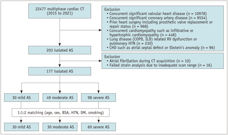
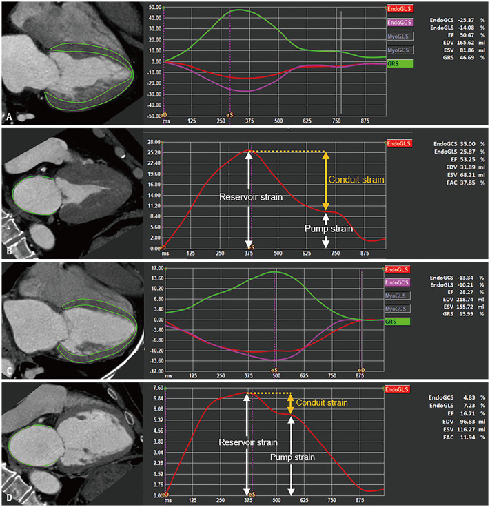
Materials and methods: This single-center retrospective study included 120 patients (median age, 76 years; 45.0% male), comprising 30, 30, and 60 patients with mild, moderate, and severe AS, respectively, all of whom underwent multiphase cardiac CT between 2015 and 2021. Patients were selected from 177 individuals who met the initial eligibility criteria, with matching for age, sex, and hypertension in a 1:1:2 ratio across the mild, moderate, and severe AS groups. Electrocardiography-gated cardiac CT images were analyzed to obtain various quantitative left ventricle (LV) and left atrium (LA) strain parameters. Statistical differences in cardiac CT-derived LV and LA strain parameters among mild, moderate, and severe AS were evaluated using the Kruskal-Wallis test, followed by post-hoc tests.
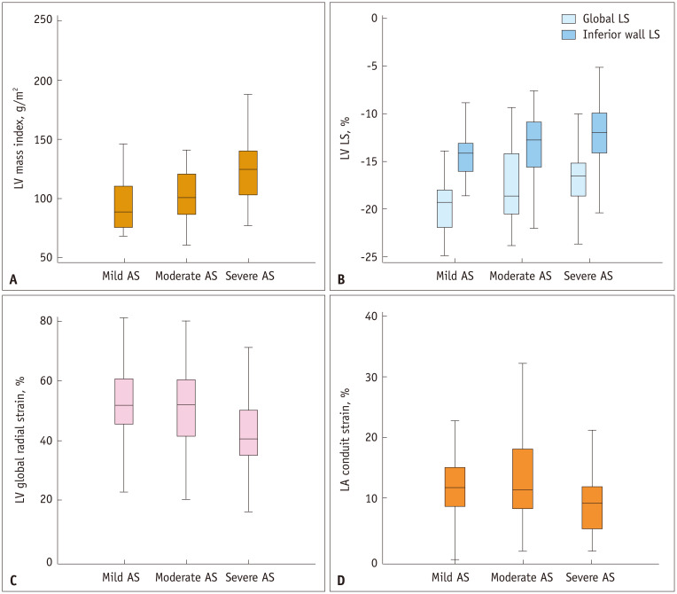
Results: The median LV global longitudinal strain differed significantly across AS severity (GLS: -19.4%, -18.2%, and -16.2% for mild, moderate, and severe AS, respectively; P < 0.001), with the absolute value decreasing as AS severity increased. Additionally, the median values of LV global circumferential strain (GCS: -29.8%, -30.8%, and -27.4%, respectively; P = 0.045), LV global radial strain (GRS: 50.1%, 50.3%, and 39.3%, respectively; P = 0.004), and LA conduit strain (11.5%, 11.2%, and 9.0%, respectively; P = 0.031) differed significantly according to AS severity, with lower absolute values observed in patients with severe AS.
Conclusion: In patients with AS, CT-derived LV and LA strains revealed changes in myocardial deformation according to AS severity. Specifically, there was a gradual decrease in the absolute value of LV GLS with increasing AS severity and initial preservation until moderate AS, followed by an eventual decrease in the absolute values of LV GCS, LV GRS, and LA conduit strain in severe AS.
期刊介绍:
The inaugural issue of the Korean J Radiol came out in March 2000. Our journal aims to produce and propagate knowledge on radiologic imaging and related sciences.
A unique feature of the articles published in the Journal will be their reflection of global trends in radiology combined with an East-Asian perspective. Geographic differences in disease prevalence will be reflected in the contents of papers, and this will serve to enrich our body of knowledge.
World''s outstanding radiologists from many countries are serving as editorial board of our journal.

 求助内容:
求助内容: 应助结果提醒方式:


