Ramy Abdelnaby, Yasmine H Ahmed, Dalia Zaafar, Mohamed Y Mahmoud, Eman Mohammed Elsaeed, Alexa Häger, Heba M A Khalil
{"title":"咽部和舌部肌肉的结构变化是阿尔茨海默病大鼠模型中吞咽困难的潜在因素。","authors":"Ramy Abdelnaby, Yasmine H Ahmed, Dalia Zaafar, Mohamed Y Mahmoud, Eman Mohammed Elsaeed, Alexa Häger, Heba M A Khalil","doi":"10.32598/bcn.2023.5719.1","DOIUrl":null,"url":null,"abstract":"<p><strong>Introduction: </strong>Alzheimer disease (AD) is a progressive neurodegenerative disease that accounts for 60% of dementia cases worldwide. Despite the lack of concrete information about the prevalence of dysphagia among AD patients, it still significantly impairs their quality of life (QoL). That outcome necessitates more investigations to understand the pathophysiology of this condition and how to manage it. In this study, we examined if AD-associated changes in pharyngeal and tongue muscles could explain dysphagia.</p><p><strong>Methods: </strong>Fourteen adult male rats were allocated into 2 groups: Group I (control) received distilled water orally, group II (AD) received aluminum chloride (AlCl<sub>3</sub>) (200 mg/kg, per os) and D-galactose (60 mg/kg, subcutaneous) daily for 45 days. Biochemical parameters were conducted, including amyloid beta-peptide (Aβ), histopathological investigation of the hippocampus, tongue, and pharynx, and immune-histochemical expression of brain glial fibrillar acidic protein (GFAP).</p><p><strong>Results: </strong>Our AD model showed marked cognitive impairment, hippocampal oxidative stress, and increased brain Aβ expression (P=0.0003) compared to controls. Dysphagia was confirmed by loss of body weight (P=0.0077) and decreased eating and drinking patterns by 25%-35% in AD versus the control group. Histopathological, immune-histochemical, and biochemical evidence, including increased levels of pharyngeal Aβ (P=0.0017), were detected in AD rats' tongue and pharyngeal muscles.</p><p><strong>Conclusion: </strong>Dysphagia in AD can result not only centrally but also due to local involvement of the tongue and pharynx. Further translational studies linking dysphagia to AD pathology will be needed.</p>","PeriodicalId":8701,"journal":{"name":"Basic and Clinical Neuroscience","volume":"15 5","pages":"671-682"},"PeriodicalIF":1.1000,"publicationDate":"2024-09-01","publicationTypes":"Journal Article","fieldsOfStudy":null,"isOpenAccess":false,"openAccessPdf":"https://www.ncbi.nlm.nih.gov/pmc/articles/PMC12198740/pdf/","citationCount":"0","resultStr":"{\"title\":\"Structural Changes in Pharyngeal and Tongue Muscles as a Potential Contributor to Dysphagia in Alzheimer Disease Rat Model.\",\"authors\":\"Ramy Abdelnaby, Yasmine H Ahmed, Dalia Zaafar, Mohamed Y Mahmoud, Eman Mohammed Elsaeed, Alexa Häger, Heba M A Khalil\",\"doi\":\"10.32598/bcn.2023.5719.1\",\"DOIUrl\":null,\"url\":null,\"abstract\":\"<p><strong>Introduction: </strong>Alzheimer disease (AD) is a progressive neurodegenerative disease that accounts for 60% of dementia cases worldwide. Despite the lack of concrete information about the prevalence of dysphagia among AD patients, it still significantly impairs their quality of life (QoL). That outcome necessitates more investigations to understand the pathophysiology of this condition and how to manage it. In this study, we examined if AD-associated changes in pharyngeal and tongue muscles could explain dysphagia.</p><p><strong>Methods: </strong>Fourteen adult male rats were allocated into 2 groups: Group I (control) received distilled water orally, group II (AD) received aluminum chloride (AlCl<sub>3</sub>) (200 mg/kg, per os) and D-galactose (60 mg/kg, subcutaneous) daily for 45 days. Biochemical parameters were conducted, including amyloid beta-peptide (Aβ), histopathological investigation of the hippocampus, tongue, and pharynx, and immune-histochemical expression of brain glial fibrillar acidic protein (GFAP).</p><p><strong>Results: </strong>Our AD model showed marked cognitive impairment, hippocampal oxidative stress, and increased brain Aβ expression (P=0.0003) compared to controls. Dysphagia was confirmed by loss of body weight (P=0.0077) and decreased eating and drinking patterns by 25%-35% in AD versus the control group. Histopathological, immune-histochemical, and biochemical evidence, including increased levels of pharyngeal Aβ (P=0.0017), were detected in AD rats' tongue and pharyngeal muscles.</p><p><strong>Conclusion: </strong>Dysphagia in AD can result not only centrally but also due to local involvement of the tongue and pharynx. Further translational studies linking dysphagia to AD pathology will be needed.</p>\",\"PeriodicalId\":8701,\"journal\":{\"name\":\"Basic and Clinical Neuroscience\",\"volume\":\"15 5\",\"pages\":\"671-682\"},\"PeriodicalIF\":1.1000,\"publicationDate\":\"2024-09-01\",\"publicationTypes\":\"Journal Article\",\"fieldsOfStudy\":null,\"isOpenAccess\":false,\"openAccessPdf\":\"https://www.ncbi.nlm.nih.gov/pmc/articles/PMC12198740/pdf/\",\"citationCount\":\"0\",\"resultStr\":null,\"platform\":\"Semanticscholar\",\"paperid\":null,\"PeriodicalName\":\"Basic and Clinical Neuroscience\",\"FirstCategoryId\":\"1085\",\"ListUrlMain\":\"https://doi.org/10.32598/bcn.2023.5719.1\",\"RegionNum\":0,\"RegionCategory\":null,\"ArticlePicture\":[],\"TitleCN\":null,\"AbstractTextCN\":null,\"PMCID\":null,\"EPubDate\":\"\",\"PubModel\":\"\",\"JCR\":\"Q4\",\"JCRName\":\"NEUROSCIENCES\",\"Score\":null,\"Total\":0}","platform":"Semanticscholar","paperid":null,"PeriodicalName":"Basic and Clinical Neuroscience","FirstCategoryId":"1085","ListUrlMain":"https://doi.org/10.32598/bcn.2023.5719.1","RegionNum":0,"RegionCategory":null,"ArticlePicture":[],"TitleCN":null,"AbstractTextCN":null,"PMCID":null,"EPubDate":"","PubModel":"","JCR":"Q4","JCRName":"NEUROSCIENCES","Score":null,"Total":0}
Structural Changes in Pharyngeal and Tongue Muscles as a Potential Contributor to Dysphagia in Alzheimer Disease Rat Model.
Introduction: Alzheimer disease (AD) is a progressive neurodegenerative disease that accounts for 60% of dementia cases worldwide. Despite the lack of concrete information about the prevalence of dysphagia among AD patients, it still significantly impairs their quality of life (QoL). That outcome necessitates more investigations to understand the pathophysiology of this condition and how to manage it. In this study, we examined if AD-associated changes in pharyngeal and tongue muscles could explain dysphagia.
Methods: Fourteen adult male rats were allocated into 2 groups: Group I (control) received distilled water orally, group II (AD) received aluminum chloride (AlCl3) (200 mg/kg, per os) and D-galactose (60 mg/kg, subcutaneous) daily for 45 days. Biochemical parameters were conducted, including amyloid beta-peptide (Aβ), histopathological investigation of the hippocampus, tongue, and pharynx, and immune-histochemical expression of brain glial fibrillar acidic protein (GFAP).
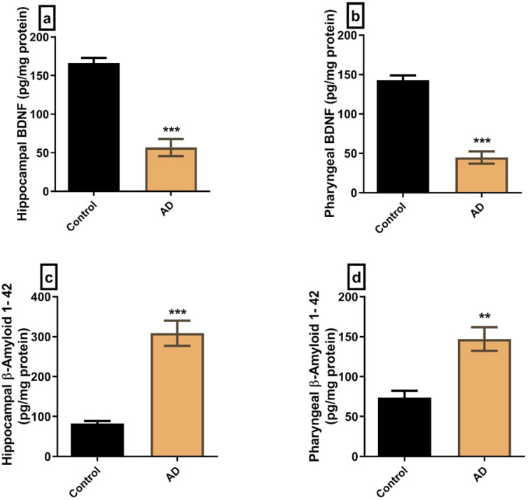
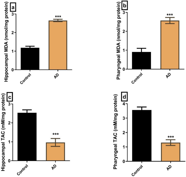
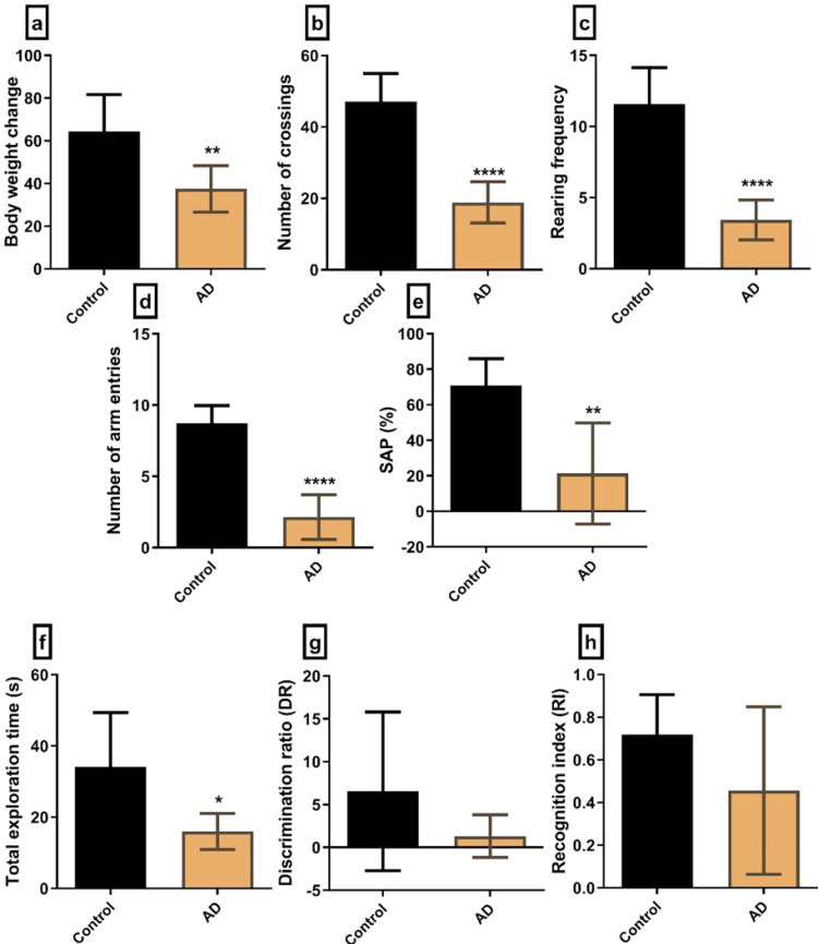
Results: Our AD model showed marked cognitive impairment, hippocampal oxidative stress, and increased brain Aβ expression (P=0.0003) compared to controls. Dysphagia was confirmed by loss of body weight (P=0.0077) and decreased eating and drinking patterns by 25%-35% in AD versus the control group. Histopathological, immune-histochemical, and biochemical evidence, including increased levels of pharyngeal Aβ (P=0.0017), were detected in AD rats' tongue and pharyngeal muscles.
Conclusion: Dysphagia in AD can result not only centrally but also due to local involvement of the tongue and pharynx. Further translational studies linking dysphagia to AD pathology will be needed.
期刊介绍:
BCN is an international multidisciplinary journal that publishes editorials, original full-length research articles, short communications, reviews, methodological papers, commentaries, perspectives and “news and reports” in the broad fields of developmental, molecular, cellular, system, computational, behavioral, cognitive, and clinical neuroscience. No area in the neural related sciences is excluded from consideration, although priority is given to studies that provide applied insights into the functioning of the nervous system. BCN aims to advance our understanding of organization and function of the nervous system in health and disease, thereby improving the diagnosis and treatment of neural-related disorders. Manuscripts submitted to BCN should describe novel results generated by experiments that were guided by clearly defined aims or hypotheses. BCN aims to provide serious ties in interdisciplinary communication, accessibility to a broad readership inside Iran and the region and also in all other international academic sites, effective peer review process, and independence from all possible non-scientific interests. BCN also tries to empower national, regional and international collaborative networks in the field of neuroscience in Iran, Middle East, Central Asia and North Africa and to be the voice of the Iranian and regional neuroscience community in the world of neuroscientists. In this way, the journal encourages submission of editorials, review papers, commentaries, methodological notes and perspectives that address this scope.
 求助内容:
求助内容: 应助结果提醒方式:


