{"title":"前列腺特异性膜抗原正电子发射断层扫描时代的全身骨扫描检测骨转移:前列腺根治术后前列腺癌患者的回顾性队列研究。","authors":"Chanikarn Poenateetai, Achiraya Teyateeti, Pawana Pusuwan, Ajalaya Teyateeti","doi":"10.22038/aojnmb.2025.82544.1582","DOIUrl":null,"url":null,"abstract":"<p><strong>Objectives: </strong>To determine the detection rate of bone metastasis on bone scan of prostate cancer patients with rising serum prostate-specific antigen (PSA) following radical prostatectomy (RP) and to identify the predictive factors associated with bone metastasis.</p><p><strong>Methods: </strong>A study was conducted in 120 patients with rising serum PSA after RP. The data collected were pre and post-RP clinical parameters, including a trigger PSA (tPSA) level that prompted the treating physician to request a bone scan and PSA doubling time (PSADT). Bone scans were classified as positive or negative in conjunction with follow-up imaging and clinical information.</p><p><strong>Results: </strong>Of 120 bone scans, 6 (5%) were positive and 114 (95%) were negative for bone metastasis. In the median tPSA ranges of <0.5, 0.5-1.0, and >1.0 ng/mL, scan positivity was 2.1%, 6.3%, and 30%, respectively. Patients with positive scans showed higher tPSA (1.228 vs 0.256 ng/mL; p=0.003) and shorter PSADT (3.5 vs 12.2 months; p=0.005) than those with negative scans. The most significant predictors of a positive bone scan were tPSA (>1 vs ≤1 ng/mL; OR 15.286, 95% CI 2.594-90.064, p=0.003) and PSADT (<6 vs ≥6 months; OR 17.333, 95% CI 1.618-185.646, p=0.018).</p><p><strong>Conclusion: </strong>The detection rate of bone metastasis on bone scans in post-RP recurrent prostate cancer patients is only 5%, but the probability is much higher with tPSA >1 ng/mL and PSADT <6 months. Given its wide accessibility in Thailand, a bone scan should remain the preferred screening test for bone metastasis, with expected positive results in patients with high or rapidly rising PSA levels.</p>","PeriodicalId":8503,"journal":{"name":"Asia Oceania Journal of Nuclear Medicine and Biology","volume":"13 2","pages":"146-155"},"PeriodicalIF":0.0000,"publicationDate":"2025-01-01","publicationTypes":"Journal Article","fieldsOfStudy":null,"isOpenAccess":false,"openAccessPdf":"https://www.ncbi.nlm.nih.gov/pmc/articles/PMC12205133/pdf/","citationCount":"0","resultStr":"{\"title\":\"Whole-Body Bone Scan for Detecting Bone Metastasis in the Prostate-Specific Membrane Antigen Positron Emission Tomography Era: A Retrospective Cohort Study of Post-Radical Prostatectomy Prostate Cancer Patients.\",\"authors\":\"Chanikarn Poenateetai, Achiraya Teyateeti, Pawana Pusuwan, Ajalaya Teyateeti\",\"doi\":\"10.22038/aojnmb.2025.82544.1582\",\"DOIUrl\":null,\"url\":null,\"abstract\":\"<p><strong>Objectives: </strong>To determine the detection rate of bone metastasis on bone scan of prostate cancer patients with rising serum prostate-specific antigen (PSA) following radical prostatectomy (RP) and to identify the predictive factors associated with bone metastasis.</p><p><strong>Methods: </strong>A study was conducted in 120 patients with rising serum PSA after RP. The data collected were pre and post-RP clinical parameters, including a trigger PSA (tPSA) level that prompted the treating physician to request a bone scan and PSA doubling time (PSADT). Bone scans were classified as positive or negative in conjunction with follow-up imaging and clinical information.</p><p><strong>Results: </strong>Of 120 bone scans, 6 (5%) were positive and 114 (95%) were negative for bone metastasis. In the median tPSA ranges of <0.5, 0.5-1.0, and >1.0 ng/mL, scan positivity was 2.1%, 6.3%, and 30%, respectively. Patients with positive scans showed higher tPSA (1.228 vs 0.256 ng/mL; p=0.003) and shorter PSADT (3.5 vs 12.2 months; p=0.005) than those with negative scans. The most significant predictors of a positive bone scan were tPSA (>1 vs ≤1 ng/mL; OR 15.286, 95% CI 2.594-90.064, p=0.003) and PSADT (<6 vs ≥6 months; OR 17.333, 95% CI 1.618-185.646, p=0.018).</p><p><strong>Conclusion: </strong>The detection rate of bone metastasis on bone scans in post-RP recurrent prostate cancer patients is only 5%, but the probability is much higher with tPSA >1 ng/mL and PSADT <6 months. Given its wide accessibility in Thailand, a bone scan should remain the preferred screening test for bone metastasis, with expected positive results in patients with high or rapidly rising PSA levels.</p>\",\"PeriodicalId\":8503,\"journal\":{\"name\":\"Asia Oceania Journal of Nuclear Medicine and Biology\",\"volume\":\"13 2\",\"pages\":\"146-155\"},\"PeriodicalIF\":0.0000,\"publicationDate\":\"2025-01-01\",\"publicationTypes\":\"Journal Article\",\"fieldsOfStudy\":null,\"isOpenAccess\":false,\"openAccessPdf\":\"https://www.ncbi.nlm.nih.gov/pmc/articles/PMC12205133/pdf/\",\"citationCount\":\"0\",\"resultStr\":null,\"platform\":\"Semanticscholar\",\"paperid\":null,\"PeriodicalName\":\"Asia Oceania Journal of Nuclear Medicine and Biology\",\"FirstCategoryId\":\"1085\",\"ListUrlMain\":\"https://doi.org/10.22038/aojnmb.2025.82544.1582\",\"RegionNum\":0,\"RegionCategory\":null,\"ArticlePicture\":[],\"TitleCN\":null,\"AbstractTextCN\":null,\"PMCID\":null,\"EPubDate\":\"\",\"PubModel\":\"\",\"JCR\":\"Q3\",\"JCRName\":\"Medicine\",\"Score\":null,\"Total\":0}","platform":"Semanticscholar","paperid":null,"PeriodicalName":"Asia Oceania Journal of Nuclear Medicine and Biology","FirstCategoryId":"1085","ListUrlMain":"https://doi.org/10.22038/aojnmb.2025.82544.1582","RegionNum":0,"RegionCategory":null,"ArticlePicture":[],"TitleCN":null,"AbstractTextCN":null,"PMCID":null,"EPubDate":"","PubModel":"","JCR":"Q3","JCRName":"Medicine","Score":null,"Total":0}
引用次数: 0
摘要
目的:探讨根治性前列腺切除术(RP)后血清前列腺特异性抗原(PSA)升高的前列腺癌患者骨扫描的骨转移检出率,并探讨骨转移的相关预测因素。方法:对120例RP术后血清PSA升高的患者进行研究。收集的数据是rp前和rp后的临床参数,包括触发PSA (tPSA)水平,促使治疗医生要求进行骨扫描和PSA倍增时间(PSADT)。结合随访影像和临床信息,将骨扫描分为阳性或阴性。结果:120例骨扫描中,骨转移阳性6例(5%),阴性114例(95%)。在tPSA中位数为1.0 ng/mL范围内,扫描阳性率分别为2.1%、6.3%和30%。扫描阳性患者tPSA升高(1.228 vs 0.256 ng/mL;p=0.003)和较短的PSADT (3.5 vs 12.2个月;P =0.005)。骨扫描阳性最显著的预测因子是tPSA (>.1 vs≤1 ng/mL;OR 15.286, 95% CI 2.594-90.064, p=0.003)和PSADT(结论:rp后复发前列腺癌患者骨扫描骨转移检出率仅为5%,但tPSA bb0.1 ng/mL和PSADT检出率高得多
Whole-Body Bone Scan for Detecting Bone Metastasis in the Prostate-Specific Membrane Antigen Positron Emission Tomography Era: A Retrospective Cohort Study of Post-Radical Prostatectomy Prostate Cancer Patients.
Objectives: To determine the detection rate of bone metastasis on bone scan of prostate cancer patients with rising serum prostate-specific antigen (PSA) following radical prostatectomy (RP) and to identify the predictive factors associated with bone metastasis.
Methods: A study was conducted in 120 patients with rising serum PSA after RP. The data collected were pre and post-RP clinical parameters, including a trigger PSA (tPSA) level that prompted the treating physician to request a bone scan and PSA doubling time (PSADT). Bone scans were classified as positive or negative in conjunction with follow-up imaging and clinical information.
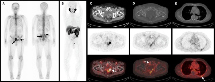
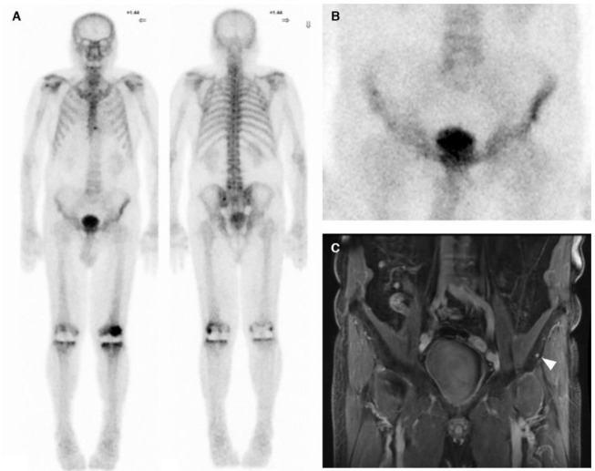
Results: Of 120 bone scans, 6 (5%) were positive and 114 (95%) were negative for bone metastasis. In the median tPSA ranges of <0.5, 0.5-1.0, and >1.0 ng/mL, scan positivity was 2.1%, 6.3%, and 30%, respectively. Patients with positive scans showed higher tPSA (1.228 vs 0.256 ng/mL; p=0.003) and shorter PSADT (3.5 vs 12.2 months; p=0.005) than those with negative scans. The most significant predictors of a positive bone scan were tPSA (>1 vs ≤1 ng/mL; OR 15.286, 95% CI 2.594-90.064, p=0.003) and PSADT (<6 vs ≥6 months; OR 17.333, 95% CI 1.618-185.646, p=0.018).
Conclusion: The detection rate of bone metastasis on bone scans in post-RP recurrent prostate cancer patients is only 5%, but the probability is much higher with tPSA >1 ng/mL and PSADT <6 months. Given its wide accessibility in Thailand, a bone scan should remain the preferred screening test for bone metastasis, with expected positive results in patients with high or rapidly rising PSA levels.

 求助内容:
求助内容: 应助结果提醒方式:


