{"title":"与传统代谢参数相比,FDG PET/CT放射组学在预测肝细胞癌微血管侵袭中的诊断性能:一项系统综述和荟萃分析。","authors":"Sang-Woo Lee, Shin Young Jeong, Seong-Jang Kim","doi":"10.1007/s12149-025-02075-y","DOIUrl":null,"url":null,"abstract":"<div><h3>Purpose</h3><p>The purpose of the current study was to evaluate the diagnostic accuracy of FDG PET/CT radiomics in predicting microvascular invasion (MVI) in hepatocellular carcinoma (HCC), and to compare it with conventional metabolic parameters of FDG PET/CT through a systematic review and meta-analysis.</p><h3>Methods</h3><p>The PubMed, EMBASE, and Cochrane databases were searched for studies evaluating the diagnostic performance of FDG PET/CT in predicting MVI in HCC patients. We calculated the pooled area under the curve (AUC) for predicting MVI using FDG PET/CT analyzed with radiomics methods and compared the results with those predicted through visual or semi-quantitative analysis. The study was conducted and registered in PROSPERO (International Prospective Register of Systematic Reviews) with the registration number CRD42023466842.</p><h3>Results</h3><p>The pooled AUC for predicting MVI from three studies (274 patients) analyzed using radiomics methods was 0.79 (95% CI; 0.75–0.84), with various model algorithms and selected features. The pooled AUC for six studies (368 patients) using visual analysis was 0.76 (95% CI; 0.73–0.80), and the pooled AUC for nine studies (661 patients) using semi-quantitative analysis was 0.80 (95% CI; 0.76–0.83). The diagnostic performance of the three analysis methods did not show a statistically significant difference.</p><h3>Conclusion</h3><p>FDG PET/CT radiomics for predicting MVI in HCC showed diagnostic performance similar to that of conventional visual or semi-quantitative analysis methods. Further large-scale multicenter studies are necessary to substantiate the diagnostic accuracy of FDG PET/CT radiomics for predicting MVI in HCC patients.</p></div>","PeriodicalId":8007,"journal":{"name":"Annals of Nuclear Medicine","volume":"39 10","pages":"1146 - 1156"},"PeriodicalIF":2.5000,"publicationDate":"2025-06-28","publicationTypes":"Journal Article","fieldsOfStudy":null,"isOpenAccess":false,"openAccessPdf":"","citationCount":"0","resultStr":"{\"title\":\"Diagnostic performance of FDG PET/CT radiomics in predicting microvascular invasion in hepatocellular carcinoma compared to conventional metabolic parameters: a systematic review and meta-analysis\",\"authors\":\"Sang-Woo Lee, Shin Young Jeong, Seong-Jang Kim\",\"doi\":\"10.1007/s12149-025-02075-y\",\"DOIUrl\":null,\"url\":null,\"abstract\":\"<div><h3>Purpose</h3><p>The purpose of the current study was to evaluate the diagnostic accuracy of FDG PET/CT radiomics in predicting microvascular invasion (MVI) in hepatocellular carcinoma (HCC), and to compare it with conventional metabolic parameters of FDG PET/CT through a systematic review and meta-analysis.</p><h3>Methods</h3><p>The PubMed, EMBASE, and Cochrane databases were searched for studies evaluating the diagnostic performance of FDG PET/CT in predicting MVI in HCC patients. We calculated the pooled area under the curve (AUC) for predicting MVI using FDG PET/CT analyzed with radiomics methods and compared the results with those predicted through visual or semi-quantitative analysis. The study was conducted and registered in PROSPERO (International Prospective Register of Systematic Reviews) with the registration number CRD42023466842.</p><h3>Results</h3><p>The pooled AUC for predicting MVI from three studies (274 patients) analyzed using radiomics methods was 0.79 (95% CI; 0.75–0.84), with various model algorithms and selected features. The pooled AUC for six studies (368 patients) using visual analysis was 0.76 (95% CI; 0.73–0.80), and the pooled AUC for nine studies (661 patients) using semi-quantitative analysis was 0.80 (95% CI; 0.76–0.83). The diagnostic performance of the three analysis methods did not show a statistically significant difference.</p><h3>Conclusion</h3><p>FDG PET/CT radiomics for predicting MVI in HCC showed diagnostic performance similar to that of conventional visual or semi-quantitative analysis methods. Further large-scale multicenter studies are necessary to substantiate the diagnostic accuracy of FDG PET/CT radiomics for predicting MVI in HCC patients.</p></div>\",\"PeriodicalId\":8007,\"journal\":{\"name\":\"Annals of Nuclear Medicine\",\"volume\":\"39 10\",\"pages\":\"1146 - 1156\"},\"PeriodicalIF\":2.5000,\"publicationDate\":\"2025-06-28\",\"publicationTypes\":\"Journal Article\",\"fieldsOfStudy\":null,\"isOpenAccess\":false,\"openAccessPdf\":\"\",\"citationCount\":\"0\",\"resultStr\":null,\"platform\":\"Semanticscholar\",\"paperid\":null,\"PeriodicalName\":\"Annals of Nuclear Medicine\",\"FirstCategoryId\":\"3\",\"ListUrlMain\":\"https://link.springer.com/article/10.1007/s12149-025-02075-y\",\"RegionNum\":4,\"RegionCategory\":\"医学\",\"ArticlePicture\":[],\"TitleCN\":null,\"AbstractTextCN\":null,\"PMCID\":null,\"EPubDate\":\"\",\"PubModel\":\"\",\"JCR\":\"Q2\",\"JCRName\":\"RADIOLOGY, NUCLEAR MEDICINE & MEDICAL IMAGING\",\"Score\":null,\"Total\":0}","platform":"Semanticscholar","paperid":null,"PeriodicalName":"Annals of Nuclear Medicine","FirstCategoryId":"3","ListUrlMain":"https://link.springer.com/article/10.1007/s12149-025-02075-y","RegionNum":4,"RegionCategory":"医学","ArticlePicture":[],"TitleCN":null,"AbstractTextCN":null,"PMCID":null,"EPubDate":"","PubModel":"","JCR":"Q2","JCRName":"RADIOLOGY, NUCLEAR MEDICINE & MEDICAL IMAGING","Score":null,"Total":0}
Diagnostic performance of FDG PET/CT radiomics in predicting microvascular invasion in hepatocellular carcinoma compared to conventional metabolic parameters: a systematic review and meta-analysis
Purpose
The purpose of the current study was to evaluate the diagnostic accuracy of FDG PET/CT radiomics in predicting microvascular invasion (MVI) in hepatocellular carcinoma (HCC), and to compare it with conventional metabolic parameters of FDG PET/CT through a systematic review and meta-analysis.
Methods
The PubMed, EMBASE, and Cochrane databases were searched for studies evaluating the diagnostic performance of FDG PET/CT in predicting MVI in HCC patients. We calculated the pooled area under the curve (AUC) for predicting MVI using FDG PET/CT analyzed with radiomics methods and compared the results with those predicted through visual or semi-quantitative analysis. The study was conducted and registered in PROSPERO (International Prospective Register of Systematic Reviews) with the registration number CRD42023466842.
Results
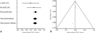
The pooled AUC for predicting MVI from three studies (274 patients) analyzed using radiomics methods was 0.79 (95% CI; 0.75–0.84), with various model algorithms and selected features. The pooled AUC for six studies (368 patients) using visual analysis was 0.76 (95% CI; 0.73–0.80), and the pooled AUC for nine studies (661 patients) using semi-quantitative analysis was 0.80 (95% CI; 0.76–0.83). The diagnostic performance of the three analysis methods did not show a statistically significant difference.
Conclusion
FDG PET/CT radiomics for predicting MVI in HCC showed diagnostic performance similar to that of conventional visual or semi-quantitative analysis methods. Further large-scale multicenter studies are necessary to substantiate the diagnostic accuracy of FDG PET/CT radiomics for predicting MVI in HCC patients.
期刊介绍:
Annals of Nuclear Medicine is an official journal of the Japanese Society of Nuclear Medicine. It develops the appropriate application of radioactive substances and stable nuclides in the field of medicine.
The journal promotes the exchange of ideas and information and research in nuclear medicine and includes the medical application of radionuclides and related subjects. It presents original articles, short communications, reviews and letters to the editor.


 求助内容:
求助内容: 应助结果提醒方式:


