Yi-Yen Tsai, Jeng-Wei Lu, Hao Yen, Chih-Chien Wang
{"title":"足跟区疼痛性血管肌瘤1例报告。","authors":"Yi-Yen Tsai, Jeng-Wei Lu, Hao Yen, Chih-Chien Wang","doi":"10.21873/invivo.14049","DOIUrl":null,"url":null,"abstract":"<p><strong>Background/aim: </strong>Angiomyomas are rare benign smooth muscle tumors originating from the tunica media of blood vessel walls, most frequently affecting the lower extremities in middle-aged women.</p><p><strong>Case report: </strong>We report the case of a 51-year-old female with a six-month history of a painful, palpable mass in the right heel. Physical examination revealed a soft, mobile, and tender subcutaneous nodule. Ultrasound imaging identified a 0.5 cm well-defined hypoechoic lesion in the subcutaneous layer, without internal blood flow, initially suspected to be an epidermoid cyst or fibrous tumor. Surgical excision of the lesion was performed, and histopathological analysis revealed a well-encapsulated tumor consisting of spindle-shaped cells with eosinophilic cytoplasm and bland nuclei, accompanied by vascular components. Immunohistochemical staining confirmed positive expression of SMA, establishing the diagnosis of angiomyoma. The patient experienced an uneventful postoperative recovery.</p><p><strong>Conclusion: </strong>This case highlights the diagnostic challenges of angiomyomas, given their nonspecific clinical presentation and imaging findings. While magnetic resonance imaging may reveal characteristic features such as strong gadolinium enhancement, definitive diagnosis relies on histopathological evaluation. Clinicians should include angiomyoma in the differential diagnosis of painful subcutaneous masses in the foot and ankle, particularly in middle-aged women. Surgical excision remains the definitive diagnostic and therapeutic approach, with low recurrence rates reported.</p>","PeriodicalId":13364,"journal":{"name":"In vivo","volume":"39 4","pages":"2485-2488"},"PeriodicalIF":1.8000,"publicationDate":"2025-07-01","publicationTypes":"Journal Article","fieldsOfStudy":null,"isOpenAccess":false,"openAccessPdf":"https://www.ncbi.nlm.nih.gov/pmc/articles/PMC12223645/pdf/","citationCount":"0","resultStr":"{\"title\":\"Painful Angiomyoma of the Heel Region: A Rare Case Report.\",\"authors\":\"Yi-Yen Tsai, Jeng-Wei Lu, Hao Yen, Chih-Chien Wang\",\"doi\":\"10.21873/invivo.14049\",\"DOIUrl\":null,\"url\":null,\"abstract\":\"<p><strong>Background/aim: </strong>Angiomyomas are rare benign smooth muscle tumors originating from the tunica media of blood vessel walls, most frequently affecting the lower extremities in middle-aged women.</p><p><strong>Case report: </strong>We report the case of a 51-year-old female with a six-month history of a painful, palpable mass in the right heel. Physical examination revealed a soft, mobile, and tender subcutaneous nodule. Ultrasound imaging identified a 0.5 cm well-defined hypoechoic lesion in the subcutaneous layer, without internal blood flow, initially suspected to be an epidermoid cyst or fibrous tumor. Surgical excision of the lesion was performed, and histopathological analysis revealed a well-encapsulated tumor consisting of spindle-shaped cells with eosinophilic cytoplasm and bland nuclei, accompanied by vascular components. Immunohistochemical staining confirmed positive expression of SMA, establishing the diagnosis of angiomyoma. The patient experienced an uneventful postoperative recovery.</p><p><strong>Conclusion: </strong>This case highlights the diagnostic challenges of angiomyomas, given their nonspecific clinical presentation and imaging findings. While magnetic resonance imaging may reveal characteristic features such as strong gadolinium enhancement, definitive diagnosis relies on histopathological evaluation. Clinicians should include angiomyoma in the differential diagnosis of painful subcutaneous masses in the foot and ankle, particularly in middle-aged women. Surgical excision remains the definitive diagnostic and therapeutic approach, with low recurrence rates reported.</p>\",\"PeriodicalId\":13364,\"journal\":{\"name\":\"In vivo\",\"volume\":\"39 4\",\"pages\":\"2485-2488\"},\"PeriodicalIF\":1.8000,\"publicationDate\":\"2025-07-01\",\"publicationTypes\":\"Journal Article\",\"fieldsOfStudy\":null,\"isOpenAccess\":false,\"openAccessPdf\":\"https://www.ncbi.nlm.nih.gov/pmc/articles/PMC12223645/pdf/\",\"citationCount\":\"0\",\"resultStr\":null,\"platform\":\"Semanticscholar\",\"paperid\":null,\"PeriodicalName\":\"In vivo\",\"FirstCategoryId\":\"3\",\"ListUrlMain\":\"https://doi.org/10.21873/invivo.14049\",\"RegionNum\":4,\"RegionCategory\":\"医学\",\"ArticlePicture\":[],\"TitleCN\":null,\"AbstractTextCN\":null,\"PMCID\":null,\"EPubDate\":\"\",\"PubModel\":\"\",\"JCR\":\"Q3\",\"JCRName\":\"MEDICINE, RESEARCH & EXPERIMENTAL\",\"Score\":null,\"Total\":0}","platform":"Semanticscholar","paperid":null,"PeriodicalName":"In vivo","FirstCategoryId":"3","ListUrlMain":"https://doi.org/10.21873/invivo.14049","RegionNum":4,"RegionCategory":"医学","ArticlePicture":[],"TitleCN":null,"AbstractTextCN":null,"PMCID":null,"EPubDate":"","PubModel":"","JCR":"Q3","JCRName":"MEDICINE, RESEARCH & EXPERIMENTAL","Score":null,"Total":0}
Painful Angiomyoma of the Heel Region: A Rare Case Report.
Background/aim: Angiomyomas are rare benign smooth muscle tumors originating from the tunica media of blood vessel walls, most frequently affecting the lower extremities in middle-aged women.
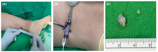
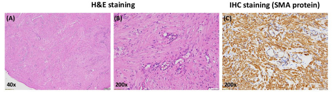
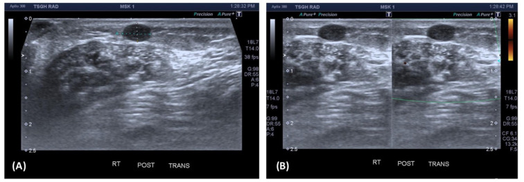
Case report: We report the case of a 51-year-old female with a six-month history of a painful, palpable mass in the right heel. Physical examination revealed a soft, mobile, and tender subcutaneous nodule. Ultrasound imaging identified a 0.5 cm well-defined hypoechoic lesion in the subcutaneous layer, without internal blood flow, initially suspected to be an epidermoid cyst or fibrous tumor. Surgical excision of the lesion was performed, and histopathological analysis revealed a well-encapsulated tumor consisting of spindle-shaped cells with eosinophilic cytoplasm and bland nuclei, accompanied by vascular components. Immunohistochemical staining confirmed positive expression of SMA, establishing the diagnosis of angiomyoma. The patient experienced an uneventful postoperative recovery.
Conclusion: This case highlights the diagnostic challenges of angiomyomas, given their nonspecific clinical presentation and imaging findings. While magnetic resonance imaging may reveal characteristic features such as strong gadolinium enhancement, definitive diagnosis relies on histopathological evaluation. Clinicians should include angiomyoma in the differential diagnosis of painful subcutaneous masses in the foot and ankle, particularly in middle-aged women. Surgical excision remains the definitive diagnostic and therapeutic approach, with low recurrence rates reported.
期刊介绍:
IN VIVO is an international peer-reviewed journal designed to bring together original high quality works and reviews on experimental and clinical biomedical research within the frames of physiology, pathology and disease management.
The topics of IN VIVO include: 1. Experimental development and application of new diagnostic and therapeutic procedures; 2. Pharmacological and toxicological evaluation of new drugs, drug combinations and drug delivery systems; 3. Clinical trials; 4. Development and characterization of models of biomedical research; 5. Cancer diagnosis and treatment; 6. Immunotherapy and vaccines; 7. Radiotherapy, Imaging; 8. Tissue engineering, Regenerative medicine; 9. Carcinogenesis.

 求助内容:
求助内容: 应助结果提醒方式:


