Esther Muñoz-Soto, Firas Elmsmari, Okba Mahmoud, José Antonio González
{"title":"病例报告:由牙根尖外表面结石样沉积引起的根尖牙周炎。","authors":"Esther Muñoz-Soto, Firas Elmsmari, Okba Mahmoud, José Antonio González","doi":"10.3389/froh.2025.1615050","DOIUrl":null,"url":null,"abstract":"<p><strong>Purpose: </strong>Herein, we report a rare case of chronic apical periodontitis associated with an extraradicular calculus-like deposit on the root apex of a mandibular left central incisor that was previously treated with root canal therapy.</p><p><strong>Case presentation: </strong>A 42-year-old man presented with persistent sinus tract formation. Despite non-surgical retreatment, the symptoms persisted, and radiographic evaluations, including cone-beam computed tomography, revealed a periapical radiolucency with radiopaque convexities in the apical third of the root. Intentional replantation (IR) was performed to allow direct clinical access for diagnosis and management. Upon extraction, a dark brown, calculus-like deposit firmly attached to the external root surface was observed. After deposit removal, root-end resection and retrograde filling were performed before replantation. Follow-ups at 3 months and 1 year revealed complete healing of the sinus tract and significant radiographic improvements. This case highlights the role of extraradicular biofilms and apical mineralized deposits in persistent periapical inflammation. Sinus tracts may facilitate mineral-ion migration and contribute to the formation of extra-radicular calculi. Mineralized biofilms may not be resolved using orthograde approaches, necessitating surgical intervention.</p><p><strong>Conclusion: </strong>IR enables thorough inspection and removal of radicular deposits, offering a minimally invasive and successful alternative to conventional apical surgery. The findings in this case are consistent with those in previous studies suggesting the usefulness of IR for managing refractory periapical lesions caused by extraradicular infections or mineralized biofilms.</p>","PeriodicalId":94016,"journal":{"name":"Frontiers in oral health","volume":"6 ","pages":"1615050"},"PeriodicalIF":3.1000,"publicationDate":"2025-06-12","publicationTypes":"Journal Article","fieldsOfStudy":null,"isOpenAccess":false,"openAccessPdf":"https://www.ncbi.nlm.nih.gov/pmc/articles/PMC12198121/pdf/","citationCount":"0","resultStr":"{\"title\":\"Case Report: Apical periodontitis due to calculus-like deposit on the external surface of the root apex.\",\"authors\":\"Esther Muñoz-Soto, Firas Elmsmari, Okba Mahmoud, José Antonio González\",\"doi\":\"10.3389/froh.2025.1615050\",\"DOIUrl\":null,\"url\":null,\"abstract\":\"<p><strong>Purpose: </strong>Herein, we report a rare case of chronic apical periodontitis associated with an extraradicular calculus-like deposit on the root apex of a mandibular left central incisor that was previously treated with root canal therapy.</p><p><strong>Case presentation: </strong>A 42-year-old man presented with persistent sinus tract formation. Despite non-surgical retreatment, the symptoms persisted, and radiographic evaluations, including cone-beam computed tomography, revealed a periapical radiolucency with radiopaque convexities in the apical third of the root. Intentional replantation (IR) was performed to allow direct clinical access for diagnosis and management. Upon extraction, a dark brown, calculus-like deposit firmly attached to the external root surface was observed. After deposit removal, root-end resection and retrograde filling were performed before replantation. Follow-ups at 3 months and 1 year revealed complete healing of the sinus tract and significant radiographic improvements. This case highlights the role of extraradicular biofilms and apical mineralized deposits in persistent periapical inflammation. Sinus tracts may facilitate mineral-ion migration and contribute to the formation of extra-radicular calculi. Mineralized biofilms may not be resolved using orthograde approaches, necessitating surgical intervention.</p><p><strong>Conclusion: </strong>IR enables thorough inspection and removal of radicular deposits, offering a minimally invasive and successful alternative to conventional apical surgery. The findings in this case are consistent with those in previous studies suggesting the usefulness of IR for managing refractory periapical lesions caused by extraradicular infections or mineralized biofilms.</p>\",\"PeriodicalId\":94016,\"journal\":{\"name\":\"Frontiers in oral health\",\"volume\":\"6 \",\"pages\":\"1615050\"},\"PeriodicalIF\":3.1000,\"publicationDate\":\"2025-06-12\",\"publicationTypes\":\"Journal Article\",\"fieldsOfStudy\":null,\"isOpenAccess\":false,\"openAccessPdf\":\"https://www.ncbi.nlm.nih.gov/pmc/articles/PMC12198121/pdf/\",\"citationCount\":\"0\",\"resultStr\":null,\"platform\":\"Semanticscholar\",\"paperid\":null,\"PeriodicalName\":\"Frontiers in oral health\",\"FirstCategoryId\":\"1085\",\"ListUrlMain\":\"https://doi.org/10.3389/froh.2025.1615050\",\"RegionNum\":0,\"RegionCategory\":null,\"ArticlePicture\":[],\"TitleCN\":null,\"AbstractTextCN\":null,\"PMCID\":null,\"EPubDate\":\"2025/1/1 0:00:00\",\"PubModel\":\"eCollection\",\"JCR\":\"Q1\",\"JCRName\":\"DENTISTRY, ORAL SURGERY & MEDICINE\",\"Score\":null,\"Total\":0}","platform":"Semanticscholar","paperid":null,"PeriodicalName":"Frontiers in oral health","FirstCategoryId":"1085","ListUrlMain":"https://doi.org/10.3389/froh.2025.1615050","RegionNum":0,"RegionCategory":null,"ArticlePicture":[],"TitleCN":null,"AbstractTextCN":null,"PMCID":null,"EPubDate":"2025/1/1 0:00:00","PubModel":"eCollection","JCR":"Q1","JCRName":"DENTISTRY, ORAL SURGERY & MEDICINE","Score":null,"Total":0}
Case Report: Apical periodontitis due to calculus-like deposit on the external surface of the root apex.
Purpose: Herein, we report a rare case of chronic apical periodontitis associated with an extraradicular calculus-like deposit on the root apex of a mandibular left central incisor that was previously treated with root canal therapy.
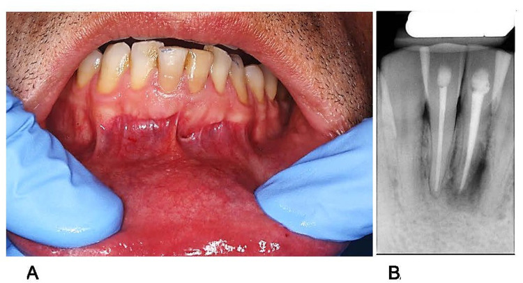
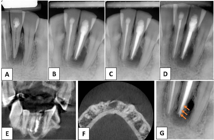
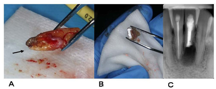
Case presentation: A 42-year-old man presented with persistent sinus tract formation. Despite non-surgical retreatment, the symptoms persisted, and radiographic evaluations, including cone-beam computed tomography, revealed a periapical radiolucency with radiopaque convexities in the apical third of the root. Intentional replantation (IR) was performed to allow direct clinical access for diagnosis and management. Upon extraction, a dark brown, calculus-like deposit firmly attached to the external root surface was observed. After deposit removal, root-end resection and retrograde filling were performed before replantation. Follow-ups at 3 months and 1 year revealed complete healing of the sinus tract and significant radiographic improvements. This case highlights the role of extraradicular biofilms and apical mineralized deposits in persistent periapical inflammation. Sinus tracts may facilitate mineral-ion migration and contribute to the formation of extra-radicular calculi. Mineralized biofilms may not be resolved using orthograde approaches, necessitating surgical intervention.
Conclusion: IR enables thorough inspection and removal of radicular deposits, offering a minimally invasive and successful alternative to conventional apical surgery. The findings in this case are consistent with those in previous studies suggesting the usefulness of IR for managing refractory periapical lesions caused by extraradicular infections or mineralized biofilms.

 求助内容:
求助内容: 应助结果提醒方式:


