Yizhen Yang, Luohai Chen, Man Liu, Xiaoxuan Lin, Sui Peng, Yubin Xie, Zhirong Zeng, Minhu Chen, Ning Zhang
{"title":"单细胞转录组显示免疫治疗在转移性胰腺血管肉瘤中的潜在作用。","authors":"Yizhen Yang, Luohai Chen, Man Liu, Xiaoxuan Lin, Sui Peng, Yubin Xie, Zhirong Zeng, Minhu Chen, Ning Zhang","doi":"10.1093/gastro/goaf046","DOIUrl":null,"url":null,"abstract":"<p><strong>Background: </strong>Pancreatic angiosarcoma is a rare and highly aggressive tumor originating from lymphatic or vascular endothelial cells, with poor prognosis and few effective treatments. In this study, we aimed to characterize the tumor ecosystem of metastatic pancreatic angiosarcoma, along with its potential treatment strategies.</p><p><strong>Methods: </strong>Single-cell RNA-sequencing and bioinformatics analysis were performed on samples obtained from one patient, including at total of 16,841 cells from pancreatic angiosarcoma liver metastasis and adjacent normal liver tissue.</p><p><strong>Results: </strong>Pancreatic angiosarcoma cells exhibited marked upregulation of nuclear factor kappa-B (NF-κB), hypoxia-inducible factor 1 (HIF-1), and myelocytomatosis oncogene (MYC) proto-oncogene signaling pathways, while presenting limited upregulation of actionable therapeutic targets except for cyclin-dependent kinase 4 (CDK4) and epidermal growth factor receptor. Several immune checkpoint genes, including cytotoxic T-lymphocyte-associated protein 4 (CTLA4), lymphocyte-activation gene 3 (LAG3), programmed cell death protein 1 (PDCD1), and cluster of differentiation 86 (CD86), were upregulated in tumor-infiltrating T cells, natural killer (NK) cells, and myeloid cells. Furthermore, intercellular interaction profiling demonstrated enhanced activity of the programmed death-ligand 1 (PD-L1) and CD86 signaling pathways within the tumor microenvironment. The gene-set scores of T/NK-cell exhaustion, regulatory T cell, and macrophage angiogenesis were significantly higher in tumor tissues compared with adjacent normal tissues. However, the phagocytosis scores of macrophages within the tumor-infiltrating region were significantly lower than those in the adjacent normal tissues.</p><p><strong>Conclusions: </strong>Our findings outlined an immunosuppressive and angiogenic tumor ecosystem in pancreatic angiosarcoma liver metastasis, suggesting that pancreatic angiosarcoma may be insensitive to most targeted therapies. Conversely, immunotherapies targeting LAG3, PD-L1, and CD86 (e.g. isatuximab, Opdualag, and abatacept) and anti-angiogenic agents may be therapeutically effective and worthy of subsequent exploration.</p>","PeriodicalId":54275,"journal":{"name":"Gastroenterology Report","volume":"13 ","pages":"goaf046"},"PeriodicalIF":4.2000,"publicationDate":"2025-06-16","publicationTypes":"Journal Article","fieldsOfStudy":null,"isOpenAccess":false,"openAccessPdf":"https://www.ncbi.nlm.nih.gov/pmc/articles/PMC12199912/pdf/","citationCount":"0","resultStr":"{\"title\":\"Single-cell transcriptomic landscape indicates the potential role of immunotherapy in metastatic pancreatic angiosarcoma.\",\"authors\":\"Yizhen Yang, Luohai Chen, Man Liu, Xiaoxuan Lin, Sui Peng, Yubin Xie, Zhirong Zeng, Minhu Chen, Ning Zhang\",\"doi\":\"10.1093/gastro/goaf046\",\"DOIUrl\":null,\"url\":null,\"abstract\":\"<p><strong>Background: </strong>Pancreatic angiosarcoma is a rare and highly aggressive tumor originating from lymphatic or vascular endothelial cells, with poor prognosis and few effective treatments. In this study, we aimed to characterize the tumor ecosystem of metastatic pancreatic angiosarcoma, along with its potential treatment strategies.</p><p><strong>Methods: </strong>Single-cell RNA-sequencing and bioinformatics analysis were performed on samples obtained from one patient, including at total of 16,841 cells from pancreatic angiosarcoma liver metastasis and adjacent normal liver tissue.</p><p><strong>Results: </strong>Pancreatic angiosarcoma cells exhibited marked upregulation of nuclear factor kappa-B (NF-κB), hypoxia-inducible factor 1 (HIF-1), and myelocytomatosis oncogene (MYC) proto-oncogene signaling pathways, while presenting limited upregulation of actionable therapeutic targets except for cyclin-dependent kinase 4 (CDK4) and epidermal growth factor receptor. Several immune checkpoint genes, including cytotoxic T-lymphocyte-associated protein 4 (CTLA4), lymphocyte-activation gene 3 (LAG3), programmed cell death protein 1 (PDCD1), and cluster of differentiation 86 (CD86), were upregulated in tumor-infiltrating T cells, natural killer (NK) cells, and myeloid cells. Furthermore, intercellular interaction profiling demonstrated enhanced activity of the programmed death-ligand 1 (PD-L1) and CD86 signaling pathways within the tumor microenvironment. The gene-set scores of T/NK-cell exhaustion, regulatory T cell, and macrophage angiogenesis were significantly higher in tumor tissues compared with adjacent normal tissues. However, the phagocytosis scores of macrophages within the tumor-infiltrating region were significantly lower than those in the adjacent normal tissues.</p><p><strong>Conclusions: </strong>Our findings outlined an immunosuppressive and angiogenic tumor ecosystem in pancreatic angiosarcoma liver metastasis, suggesting that pancreatic angiosarcoma may be insensitive to most targeted therapies. Conversely, immunotherapies targeting LAG3, PD-L1, and CD86 (e.g. isatuximab, Opdualag, and abatacept) and anti-angiogenic agents may be therapeutically effective and worthy of subsequent exploration.</p>\",\"PeriodicalId\":54275,\"journal\":{\"name\":\"Gastroenterology Report\",\"volume\":\"13 \",\"pages\":\"goaf046\"},\"PeriodicalIF\":4.2000,\"publicationDate\":\"2025-06-16\",\"publicationTypes\":\"Journal Article\",\"fieldsOfStudy\":null,\"isOpenAccess\":false,\"openAccessPdf\":\"https://www.ncbi.nlm.nih.gov/pmc/articles/PMC12199912/pdf/\",\"citationCount\":\"0\",\"resultStr\":null,\"platform\":\"Semanticscholar\",\"paperid\":null,\"PeriodicalName\":\"Gastroenterology Report\",\"FirstCategoryId\":\"3\",\"ListUrlMain\":\"https://doi.org/10.1093/gastro/goaf046\",\"RegionNum\":3,\"RegionCategory\":\"医学\",\"ArticlePicture\":[],\"TitleCN\":null,\"AbstractTextCN\":null,\"PMCID\":null,\"EPubDate\":\"2025/1/1 0:00:00\",\"PubModel\":\"eCollection\",\"JCR\":\"Q2\",\"JCRName\":\"GASTROENTEROLOGY & HEPATOLOGY\",\"Score\":null,\"Total\":0}","platform":"Semanticscholar","paperid":null,"PeriodicalName":"Gastroenterology Report","FirstCategoryId":"3","ListUrlMain":"https://doi.org/10.1093/gastro/goaf046","RegionNum":3,"RegionCategory":"医学","ArticlePicture":[],"TitleCN":null,"AbstractTextCN":null,"PMCID":null,"EPubDate":"2025/1/1 0:00:00","PubModel":"eCollection","JCR":"Q2","JCRName":"GASTROENTEROLOGY & HEPATOLOGY","Score":null,"Total":0}
Single-cell transcriptomic landscape indicates the potential role of immunotherapy in metastatic pancreatic angiosarcoma.
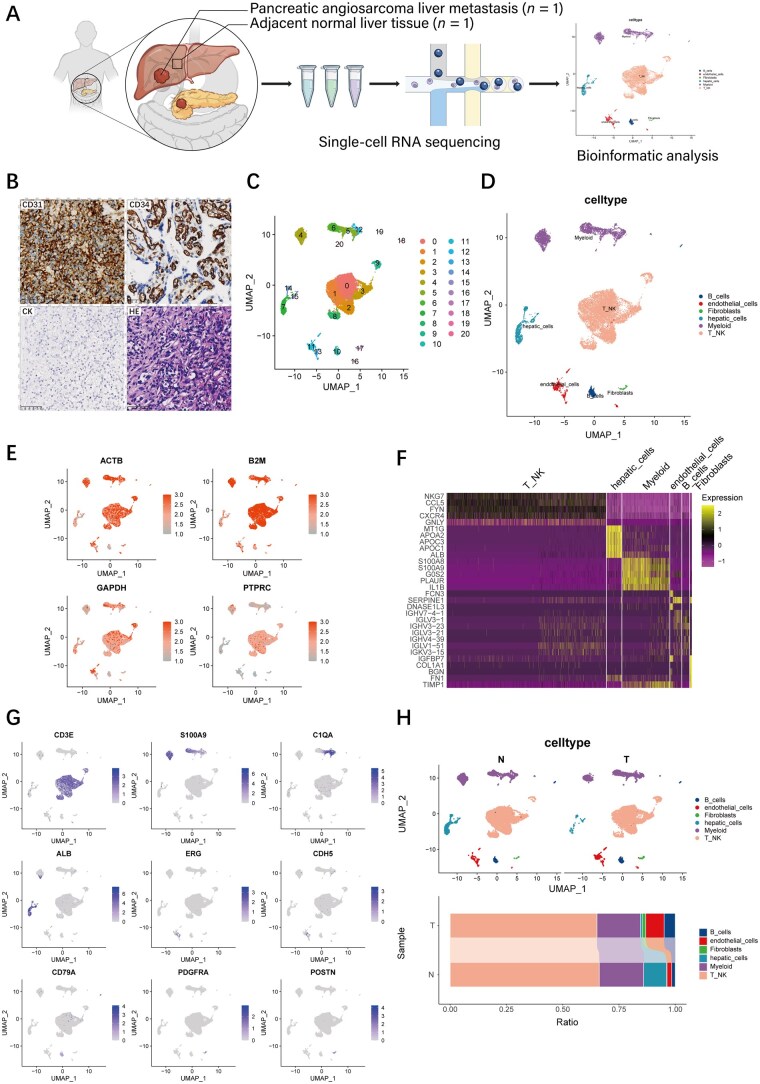
Background: Pancreatic angiosarcoma is a rare and highly aggressive tumor originating from lymphatic or vascular endothelial cells, with poor prognosis and few effective treatments. In this study, we aimed to characterize the tumor ecosystem of metastatic pancreatic angiosarcoma, along with its potential treatment strategies.
Methods: Single-cell RNA-sequencing and bioinformatics analysis were performed on samples obtained from one patient, including at total of 16,841 cells from pancreatic angiosarcoma liver metastasis and adjacent normal liver tissue.
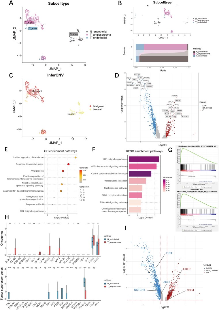
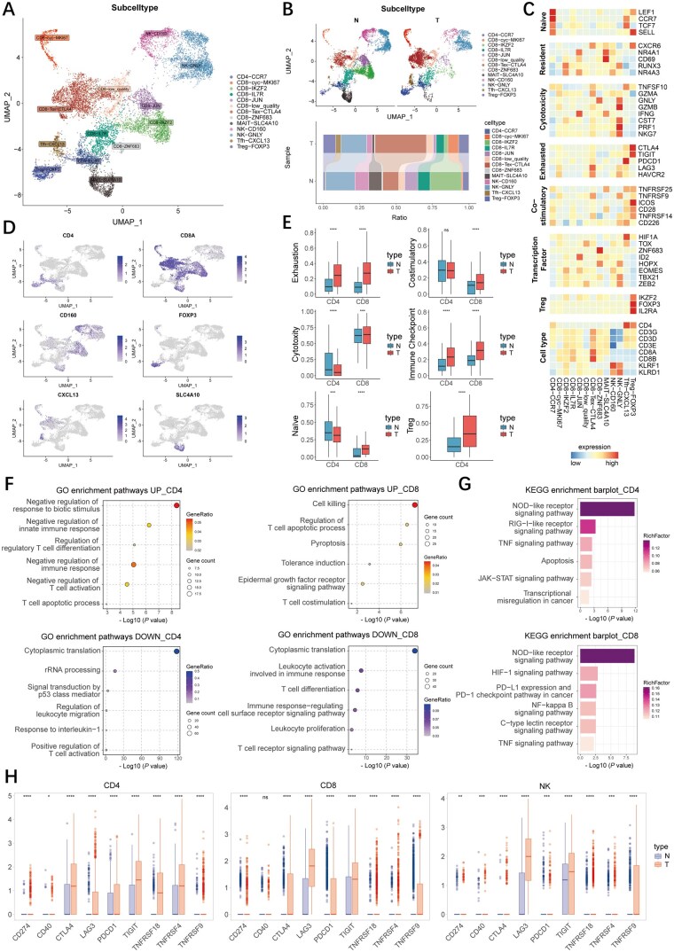
Results: Pancreatic angiosarcoma cells exhibited marked upregulation of nuclear factor kappa-B (NF-κB), hypoxia-inducible factor 1 (HIF-1), and myelocytomatosis oncogene (MYC) proto-oncogene signaling pathways, while presenting limited upregulation of actionable therapeutic targets except for cyclin-dependent kinase 4 (CDK4) and epidermal growth factor receptor. Several immune checkpoint genes, including cytotoxic T-lymphocyte-associated protein 4 (CTLA4), lymphocyte-activation gene 3 (LAG3), programmed cell death protein 1 (PDCD1), and cluster of differentiation 86 (CD86), were upregulated in tumor-infiltrating T cells, natural killer (NK) cells, and myeloid cells. Furthermore, intercellular interaction profiling demonstrated enhanced activity of the programmed death-ligand 1 (PD-L1) and CD86 signaling pathways within the tumor microenvironment. The gene-set scores of T/NK-cell exhaustion, regulatory T cell, and macrophage angiogenesis were significantly higher in tumor tissues compared with adjacent normal tissues. However, the phagocytosis scores of macrophages within the tumor-infiltrating region were significantly lower than those in the adjacent normal tissues.
Conclusions: Our findings outlined an immunosuppressive and angiogenic tumor ecosystem in pancreatic angiosarcoma liver metastasis, suggesting that pancreatic angiosarcoma may be insensitive to most targeted therapies. Conversely, immunotherapies targeting LAG3, PD-L1, and CD86 (e.g. isatuximab, Opdualag, and abatacept) and anti-angiogenic agents may be therapeutically effective and worthy of subsequent exploration.
期刊介绍:
Gastroenterology Report is an international fully open access (OA) online only journal, covering all areas related to gastrointestinal sciences, including studies of the alimentary tract, liver, biliary, pancreas, enteral nutrition and related fields. The journal aims to publish high quality research articles on both basic and clinical gastroenterology, authoritative reviews that bring together new advances in the field, as well as commentaries and highlight pieces that provide expert analysis of topical issues.
 求助内容:
求助内容: 应助结果提醒方式:


