{"title":"超声技术与磁共振成像质子密度脂肪分数测量肝纤维化儿童肝脏脂肪量的比较。","authors":"Sabriye Gulcin Bozbeyoglu, Murat Asik","doi":"10.4274/MMJ.galenos.2025.34017","DOIUrl":null,"url":null,"abstract":"<p><strong>Objective: </strong>The aim of this study is to demonstrate the reliability of Quantitative Ultrasound (QUS) in assessing liver fat content in children, using magnetic resonance imaging proton density fat fraction (MRI-PDFF) values as a reference, and to determine threshold values for QUS in grading hepatosteatosis.</p><p><strong>Methods: </strong>The study group consisted of pediatric patients under 18 years of age without known liver disease who volunteered to participate. All patients underwent MRI-PDFF scanning, and QUS imaging was performed using the tissue attenuation imaging (TAI) ve tissue scatter distribution imaging (TSI) tools. The cut-off values for MRI-PDFF were set at ≥5%, ≥16.3%, and ≥21.7%, corresponding to mild, moderate, and severe steatosis, respectively. The diagnostic performance of TAI and TSI in detecting various degrees of hepatic steatosis was evaluated using the area under the ROC (AUROC) curves.</p><p><strong>Results: </strong>The frequencies of hepatosteatosis grading were as follows: S1: 19 (37%), S2: 5 (10%), S3: 22 (43%). The AUROCs for TAI and TSI tools in detecting QUS measurements (MRI PDFF ≥5%) were 0.95 [95% confidence interval (CI): 0.91-0.99] (p< 0.001) and 0.96 (95% CI: 0.93-0.99) (p<0.001), respectively. For distinguishing different degrees of steatosis, TAI showed values of 0.75, 0.86, and 0.96 dB/cm/MHz, corresponding to sensitivities of 88%, 88%, and 100%, respectively, while TSI showed values of 92.44, 96.64, and 99.45, with sensitivities of 90%, 92%, and 91.7%. The correlation test between QUS measurements [TAI, TSI, EzHRI (Hepato-Renal Index with Automated regions of interest Recommendation)] and MR-PDFF indicated a concordance in TAI and TSI values, but not with EzHR.</p><p><strong>Conclusions: </strong>The TAI and TSI tools can accurately measure liver fat content and can be used reliably in children for the assessment and grading of hepatosteatosis.</p>","PeriodicalId":37427,"journal":{"name":"Medeniyet medical journal","volume":"40 2","pages":"46-52"},"PeriodicalIF":1.1000,"publicationDate":"2025-06-26","publicationTypes":"Journal Article","fieldsOfStudy":null,"isOpenAccess":false,"openAccessPdf":"https://www.ncbi.nlm.nih.gov/pmc/articles/PMC12203445/pdf/","citationCount":"0","resultStr":"{\"title\":\"Comparison of Ultrasound-Based Techniques and Magnetic Resonance Imaging Proton Density Fat Fraction in Measuring the Amount of Hepatic Fat in Children with Hepatosteatosis.\",\"authors\":\"Sabriye Gulcin Bozbeyoglu, Murat Asik\",\"doi\":\"10.4274/MMJ.galenos.2025.34017\",\"DOIUrl\":null,\"url\":null,\"abstract\":\"<p><strong>Objective: </strong>The aim of this study is to demonstrate the reliability of Quantitative Ultrasound (QUS) in assessing liver fat content in children, using magnetic resonance imaging proton density fat fraction (MRI-PDFF) values as a reference, and to determine threshold values for QUS in grading hepatosteatosis.</p><p><strong>Methods: </strong>The study group consisted of pediatric patients under 18 years of age without known liver disease who volunteered to participate. All patients underwent MRI-PDFF scanning, and QUS imaging was performed using the tissue attenuation imaging (TAI) ve tissue scatter distribution imaging (TSI) tools. The cut-off values for MRI-PDFF were set at ≥5%, ≥16.3%, and ≥21.7%, corresponding to mild, moderate, and severe steatosis, respectively. The diagnostic performance of TAI and TSI in detecting various degrees of hepatic steatosis was evaluated using the area under the ROC (AUROC) curves.</p><p><strong>Results: </strong>The frequencies of hepatosteatosis grading were as follows: S1: 19 (37%), S2: 5 (10%), S3: 22 (43%). The AUROCs for TAI and TSI tools in detecting QUS measurements (MRI PDFF ≥5%) were 0.95 [95% confidence interval (CI): 0.91-0.99] (p< 0.001) and 0.96 (95% CI: 0.93-0.99) (p<0.001), respectively. For distinguishing different degrees of steatosis, TAI showed values of 0.75, 0.86, and 0.96 dB/cm/MHz, corresponding to sensitivities of 88%, 88%, and 100%, respectively, while TSI showed values of 92.44, 96.64, and 99.45, with sensitivities of 90%, 92%, and 91.7%. The correlation test between QUS measurements [TAI, TSI, EzHRI (Hepato-Renal Index with Automated regions of interest Recommendation)] and MR-PDFF indicated a concordance in TAI and TSI values, but not with EzHR.</p><p><strong>Conclusions: </strong>The TAI and TSI tools can accurately measure liver fat content and can be used reliably in children for the assessment and grading of hepatosteatosis.</p>\",\"PeriodicalId\":37427,\"journal\":{\"name\":\"Medeniyet medical journal\",\"volume\":\"40 2\",\"pages\":\"46-52\"},\"PeriodicalIF\":1.1000,\"publicationDate\":\"2025-06-26\",\"publicationTypes\":\"Journal Article\",\"fieldsOfStudy\":null,\"isOpenAccess\":false,\"openAccessPdf\":\"https://www.ncbi.nlm.nih.gov/pmc/articles/PMC12203445/pdf/\",\"citationCount\":\"0\",\"resultStr\":null,\"platform\":\"Semanticscholar\",\"paperid\":null,\"PeriodicalName\":\"Medeniyet medical journal\",\"FirstCategoryId\":\"1085\",\"ListUrlMain\":\"https://doi.org/10.4274/MMJ.galenos.2025.34017\",\"RegionNum\":0,\"RegionCategory\":null,\"ArticlePicture\":[],\"TitleCN\":null,\"AbstractTextCN\":null,\"PMCID\":null,\"EPubDate\":\"\",\"PubModel\":\"\",\"JCR\":\"Q2\",\"JCRName\":\"MEDICINE, GENERAL & INTERNAL\",\"Score\":null,\"Total\":0}","platform":"Semanticscholar","paperid":null,"PeriodicalName":"Medeniyet medical journal","FirstCategoryId":"1085","ListUrlMain":"https://doi.org/10.4274/MMJ.galenos.2025.34017","RegionNum":0,"RegionCategory":null,"ArticlePicture":[],"TitleCN":null,"AbstractTextCN":null,"PMCID":null,"EPubDate":"","PubModel":"","JCR":"Q2","JCRName":"MEDICINE, GENERAL & INTERNAL","Score":null,"Total":0}
Comparison of Ultrasound-Based Techniques and Magnetic Resonance Imaging Proton Density Fat Fraction in Measuring the Amount of Hepatic Fat in Children with Hepatosteatosis.
Objective: The aim of this study is to demonstrate the reliability of Quantitative Ultrasound (QUS) in assessing liver fat content in children, using magnetic resonance imaging proton density fat fraction (MRI-PDFF) values as a reference, and to determine threshold values for QUS in grading hepatosteatosis.
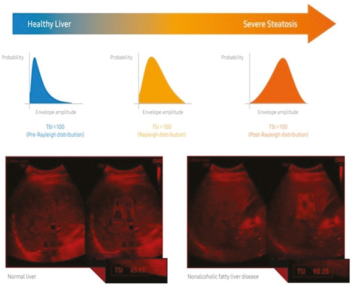
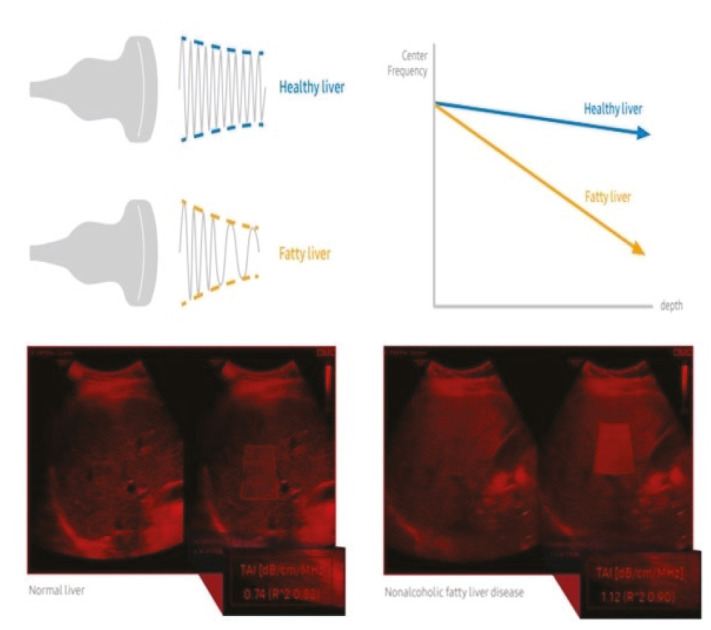
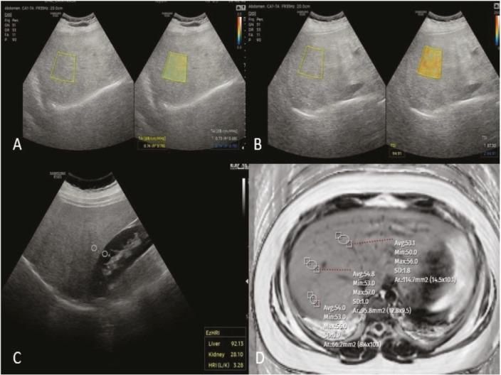
Methods: The study group consisted of pediatric patients under 18 years of age without known liver disease who volunteered to participate. All patients underwent MRI-PDFF scanning, and QUS imaging was performed using the tissue attenuation imaging (TAI) ve tissue scatter distribution imaging (TSI) tools. The cut-off values for MRI-PDFF were set at ≥5%, ≥16.3%, and ≥21.7%, corresponding to mild, moderate, and severe steatosis, respectively. The diagnostic performance of TAI and TSI in detecting various degrees of hepatic steatosis was evaluated using the area under the ROC (AUROC) curves.
Results: The frequencies of hepatosteatosis grading were as follows: S1: 19 (37%), S2: 5 (10%), S3: 22 (43%). The AUROCs for TAI and TSI tools in detecting QUS measurements (MRI PDFF ≥5%) were 0.95 [95% confidence interval (CI): 0.91-0.99] (p< 0.001) and 0.96 (95% CI: 0.93-0.99) (p<0.001), respectively. For distinguishing different degrees of steatosis, TAI showed values of 0.75, 0.86, and 0.96 dB/cm/MHz, corresponding to sensitivities of 88%, 88%, and 100%, respectively, while TSI showed values of 92.44, 96.64, and 99.45, with sensitivities of 90%, 92%, and 91.7%. The correlation test between QUS measurements [TAI, TSI, EzHRI (Hepato-Renal Index with Automated regions of interest Recommendation)] and MR-PDFF indicated a concordance in TAI and TSI values, but not with EzHR.
Conclusions: The TAI and TSI tools can accurately measure liver fat content and can be used reliably in children for the assessment and grading of hepatosteatosis.
期刊介绍:
The Medeniyet Medical Journal (Medeniyet Med J) is an open access, peer-reviewed, and scientific journal of Istanbul Medeniyet University Faculty of Medicine on various academic disciplines in medicine, which is published in English four times a year, in March, June, September, and December by a group of academics. Medeniyet Medical Journal is the continuation of Göztepe Medical Journal (ISSN: 1300-526X) which was started publishing in 1985. It changed the name as Medeniyet Medical Journal in 2015. Submission and publication are free of charge. No fees are asked from the authors for evaluation or publication process. All published articles are available online in the journal website (www.medeniyetmedicaljournal.org) without any fee. The journal publishes intradisciplinary or interdisciplinary clinical, experimental, and basic researches as well as original case reports, reviews, invited reviews, or letters to the editor, Being published since 1985, the Medeniyet Med J recognizes that the best science should lead to better lives based on the fact that the medicine should serve to the needs of society, and knowledge should transform society. The journal aims to address current issues at both national and international levels, start debates, and exert an influence on decision-makers all over the world by integrating science in everyday life. Medeniyet Med J is committed to serve the public and influence people’s lives in a positive way by making science widely accessible. Believing that the only goal is improving lives, and research has an impact on people’s lives, we select the best research papers in line with this goal.

 求助内容:
求助内容: 应助结果提醒方式:


