{"title":"鼻中隔长形皮瓣的解剖学基础。","authors":"Tor Chiu","doi":"10.14639/0392-100X-N2159","DOIUrl":null,"url":null,"abstract":"<p><strong>Objective: </strong>The aim of this study was to elucidate the anatomical factors influencing elongation of the pedicle of the nasoseptal flap (NSF).</p><p><strong>Methods: </strong>Dissections were conducted on tissue blocks taken from 61 embalmed half-heads. In the first group, 36 were microdissected to delineate the configuration of the third part of the maxillary artery (MA). In the second group, 25 other specimens were dissected using endoscopic instruments to determine the tethering points limiting the mobility of the NSF and to document the surgical manoeuvres required for release.</p><p><strong>Results: </strong>The MA is coiled in the pterygopalatine fossa (PPF) into a variety of configurations that can be classified into single-looped (SL) or double-looped (DL) forms depending on the number of vessel loops. Up to 4.7 ± 0.5 cm of NSF pedicle elongation is possible in the more common (80.8%) DL forms. Elongation is limited to 2.6 ± 0.5 cm in the SL form; greater palatine artery transection is more important to pedicle elongation in the SL form. Posterior branches such as the pharyngeal and vidian arteries may hinder pedicle elongation.</p><p><strong>Conclusions: </strong>The pedicle of the NSF may be elongated by up to 5 cm, greatly increasing its potential utility. The vessel length available for elongation is determined by the extent of MA looping, while the branch configuration, and particularly the origins of the posterior branches, determine how difficult the process will be.</p>","PeriodicalId":520544,"journal":{"name":"Acta otorhinolaryngologica Italica : organo ufficiale della Societa italiana di otorinolaringologia e chirurgia cervico-facciale","volume":"45 3","pages":"192-199"},"PeriodicalIF":0.0000,"publicationDate":"2025-06-01","publicationTypes":"Journal Article","fieldsOfStudy":null,"isOpenAccess":false,"openAccessPdf":"https://www.ncbi.nlm.nih.gov/pmc/articles/PMC12201921/pdf/","citationCount":"0","resultStr":"{\"title\":\"Anatomical basis of the elongated nasoseptal flap.\",\"authors\":\"Tor Chiu\",\"doi\":\"10.14639/0392-100X-N2159\",\"DOIUrl\":null,\"url\":null,\"abstract\":\"<p><strong>Objective: </strong>The aim of this study was to elucidate the anatomical factors influencing elongation of the pedicle of the nasoseptal flap (NSF).</p><p><strong>Methods: </strong>Dissections were conducted on tissue blocks taken from 61 embalmed half-heads. In the first group, 36 were microdissected to delineate the configuration of the third part of the maxillary artery (MA). In the second group, 25 other specimens were dissected using endoscopic instruments to determine the tethering points limiting the mobility of the NSF and to document the surgical manoeuvres required for release.</p><p><strong>Results: </strong>The MA is coiled in the pterygopalatine fossa (PPF) into a variety of configurations that can be classified into single-looped (SL) or double-looped (DL) forms depending on the number of vessel loops. Up to 4.7 ± 0.5 cm of NSF pedicle elongation is possible in the more common (80.8%) DL forms. Elongation is limited to 2.6 ± 0.5 cm in the SL form; greater palatine artery transection is more important to pedicle elongation in the SL form. Posterior branches such as the pharyngeal and vidian arteries may hinder pedicle elongation.</p><p><strong>Conclusions: </strong>The pedicle of the NSF may be elongated by up to 5 cm, greatly increasing its potential utility. The vessel length available for elongation is determined by the extent of MA looping, while the branch configuration, and particularly the origins of the posterior branches, determine how difficult the process will be.</p>\",\"PeriodicalId\":520544,\"journal\":{\"name\":\"Acta otorhinolaryngologica Italica : organo ufficiale della Societa italiana di otorinolaringologia e chirurgia cervico-facciale\",\"volume\":\"45 3\",\"pages\":\"192-199\"},\"PeriodicalIF\":0.0000,\"publicationDate\":\"2025-06-01\",\"publicationTypes\":\"Journal Article\",\"fieldsOfStudy\":null,\"isOpenAccess\":false,\"openAccessPdf\":\"https://www.ncbi.nlm.nih.gov/pmc/articles/PMC12201921/pdf/\",\"citationCount\":\"0\",\"resultStr\":null,\"platform\":\"Semanticscholar\",\"paperid\":null,\"PeriodicalName\":\"Acta otorhinolaryngologica Italica : organo ufficiale della Societa italiana di otorinolaringologia e chirurgia cervico-facciale\",\"FirstCategoryId\":\"1085\",\"ListUrlMain\":\"https://doi.org/10.14639/0392-100X-N2159\",\"RegionNum\":0,\"RegionCategory\":null,\"ArticlePicture\":[],\"TitleCN\":null,\"AbstractTextCN\":null,\"PMCID\":null,\"EPubDate\":\"\",\"PubModel\":\"\",\"JCR\":\"\",\"JCRName\":\"\",\"Score\":null,\"Total\":0}","platform":"Semanticscholar","paperid":null,"PeriodicalName":"Acta otorhinolaryngologica Italica : organo ufficiale della Societa italiana di otorinolaringologia e chirurgia cervico-facciale","FirstCategoryId":"1085","ListUrlMain":"https://doi.org/10.14639/0392-100X-N2159","RegionNum":0,"RegionCategory":null,"ArticlePicture":[],"TitleCN":null,"AbstractTextCN":null,"PMCID":null,"EPubDate":"","PubModel":"","JCR":"","JCRName":"","Score":null,"Total":0}
Anatomical basis of the elongated nasoseptal flap.
Objective: The aim of this study was to elucidate the anatomical factors influencing elongation of the pedicle of the nasoseptal flap (NSF).
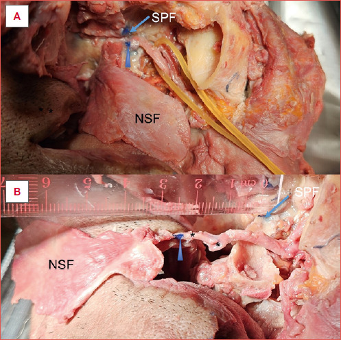
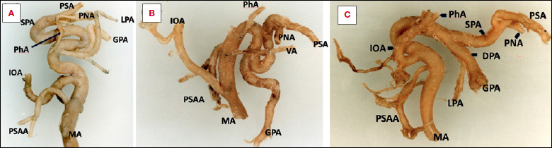
Methods: Dissections were conducted on tissue blocks taken from 61 embalmed half-heads. In the first group, 36 were microdissected to delineate the configuration of the third part of the maxillary artery (MA). In the second group, 25 other specimens were dissected using endoscopic instruments to determine the tethering points limiting the mobility of the NSF and to document the surgical manoeuvres required for release.
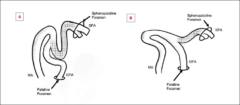
Results: The MA is coiled in the pterygopalatine fossa (PPF) into a variety of configurations that can be classified into single-looped (SL) or double-looped (DL) forms depending on the number of vessel loops. Up to 4.7 ± 0.5 cm of NSF pedicle elongation is possible in the more common (80.8%) DL forms. Elongation is limited to 2.6 ± 0.5 cm in the SL form; greater palatine artery transection is more important to pedicle elongation in the SL form. Posterior branches such as the pharyngeal and vidian arteries may hinder pedicle elongation.
Conclusions: The pedicle of the NSF may be elongated by up to 5 cm, greatly increasing its potential utility. The vessel length available for elongation is determined by the extent of MA looping, while the branch configuration, and particularly the origins of the posterior branches, determine how difficult the process will be.

 求助内容:
求助内容: 应助结果提醒方式:


