Massimo Cesareo, Alessio Martucci, Roberta Bovenzi, Marco Lombardo, Francesca Pistoia, Vittoria Carla D'Agostino, Alessandro Stefani, Carlo Nucci, Nicola Biagio Mercuri, Maria Albanese
{"title":"评估抗cgrp单克隆抗体对偏头痛患者视网膜特征的影响:一项回顾性光学相干断层扫描研究。","authors":"Massimo Cesareo, Alessio Martucci, Roberta Bovenzi, Marco Lombardo, Francesca Pistoia, Vittoria Carla D'Agostino, Alessandro Stefani, Carlo Nucci, Nicola Biagio Mercuri, Maria Albanese","doi":"10.1177/17562864251347277","DOIUrl":null,"url":null,"abstract":"<p><strong>Background: </strong>Migraine is a disabling neurovascular disorder characterized by recurrent attacks that lead to extracranial and visual involvement. Several studies have investigated the retinal vasculature features in individuals with migraine, but there have been conflicting results.</p><p><strong>Objective: </strong>To evaluate retinal structure in migraine patients before (T0) and after 6-month therapy (T1) with anti-calcitonin gene-related peptide (CGRP) monoclonal antibodies (mAbs), using optical coherence tomography (OCT) imaging.</p><p><strong>Design: </strong>A case-control and longitudinal study was conducted between January 2021 and December 2023, including 20 eyes from 10 healthy controls (HCs) and 32 eyes of 16 patients with migraine and treated with anti-CGRP mAbs according to AIFA criteria.</p><p><strong>Methods: </strong>Patients underwent OCT angiography (OCT-A) to assess retinal vessel density (VD) and spectral-domain OCT (SD-OCT) to evaluate central retinal thickness, macular structure, and peripapillary retinal nerve fiber layer thickness. These parameters were assessed in both groups at T0 and again after 6 months (T1) as part of routine clinical care.</p><p><strong>Results: </strong>All migraineurs exhibited a significant reduction in disease disability at T1, as assessed by clinical parameters. OCT data analysis revealed that individuals with migraine showed a significant increase in temporal retinal nerve fiber layer (RNFL) thickness and a reduction in nasal RNFL thickness compared to HCs. No differences in retinal circulation were observed between the groups at baseline. At T1, RNFL thickness remained sustained in the superior temporal sector, while the percentage VD of the superficial capillary plexus and radial peripapillary capillary significantly increased in the nasal perifoveal, inferior temporal, and hemi-inferior subregions.</p><p><strong>Conclusion: </strong>Our study suggests that specific retinal structural changes could precede vascular dysfunction in migraine and can be detected early by combining SD-OCT and OCT-A acquisitions. Short-term treatment with anti-CGRP mAbs may exert neuroprotective effects, potentially preventing permanent ocular damage.</p><p><strong>Trial registration: </strong>EyeHEAD Study (Trial registration number AIFA July/2024: IT 1735, www.aifa.gov.it/registro-studi-osservazionali).</p>","PeriodicalId":22980,"journal":{"name":"Therapeutic Advances in Neurological Disorders","volume":"18 ","pages":"17562864251347277"},"PeriodicalIF":4.1000,"publicationDate":"2025-06-24","publicationTypes":"Journal Article","fieldsOfStudy":null,"isOpenAccess":false,"openAccessPdf":"https://www.ncbi.nlm.nih.gov/pmc/articles/PMC12188072/pdf/","citationCount":"0","resultStr":"{\"title\":\"Evaluating the impact of anti-CGRP monoclonal antibodies on retinal features in migraine patients: a retrospective optical coherence tomography study.\",\"authors\":\"Massimo Cesareo, Alessio Martucci, Roberta Bovenzi, Marco Lombardo, Francesca Pistoia, Vittoria Carla D'Agostino, Alessandro Stefani, Carlo Nucci, Nicola Biagio Mercuri, Maria Albanese\",\"doi\":\"10.1177/17562864251347277\",\"DOIUrl\":null,\"url\":null,\"abstract\":\"<p><strong>Background: </strong>Migraine is a disabling neurovascular disorder characterized by recurrent attacks that lead to extracranial and visual involvement. Several studies have investigated the retinal vasculature features in individuals with migraine, but there have been conflicting results.</p><p><strong>Objective: </strong>To evaluate retinal structure in migraine patients before (T0) and after 6-month therapy (T1) with anti-calcitonin gene-related peptide (CGRP) monoclonal antibodies (mAbs), using optical coherence tomography (OCT) imaging.</p><p><strong>Design: </strong>A case-control and longitudinal study was conducted between January 2021 and December 2023, including 20 eyes from 10 healthy controls (HCs) and 32 eyes of 16 patients with migraine and treated with anti-CGRP mAbs according to AIFA criteria.</p><p><strong>Methods: </strong>Patients underwent OCT angiography (OCT-A) to assess retinal vessel density (VD) and spectral-domain OCT (SD-OCT) to evaluate central retinal thickness, macular structure, and peripapillary retinal nerve fiber layer thickness. These parameters were assessed in both groups at T0 and again after 6 months (T1) as part of routine clinical care.</p><p><strong>Results: </strong>All migraineurs exhibited a significant reduction in disease disability at T1, as assessed by clinical parameters. OCT data analysis revealed that individuals with migraine showed a significant increase in temporal retinal nerve fiber layer (RNFL) thickness and a reduction in nasal RNFL thickness compared to HCs. No differences in retinal circulation were observed between the groups at baseline. At T1, RNFL thickness remained sustained in the superior temporal sector, while the percentage VD of the superficial capillary plexus and radial peripapillary capillary significantly increased in the nasal perifoveal, inferior temporal, and hemi-inferior subregions.</p><p><strong>Conclusion: </strong>Our study suggests that specific retinal structural changes could precede vascular dysfunction in migraine and can be detected early by combining SD-OCT and OCT-A acquisitions. Short-term treatment with anti-CGRP mAbs may exert neuroprotective effects, potentially preventing permanent ocular damage.</p><p><strong>Trial registration: </strong>EyeHEAD Study (Trial registration number AIFA July/2024: IT 1735, www.aifa.gov.it/registro-studi-osservazionali).</p>\",\"PeriodicalId\":22980,\"journal\":{\"name\":\"Therapeutic Advances in Neurological Disorders\",\"volume\":\"18 \",\"pages\":\"17562864251347277\"},\"PeriodicalIF\":4.1000,\"publicationDate\":\"2025-06-24\",\"publicationTypes\":\"Journal Article\",\"fieldsOfStudy\":null,\"isOpenAccess\":false,\"openAccessPdf\":\"https://www.ncbi.nlm.nih.gov/pmc/articles/PMC12188072/pdf/\",\"citationCount\":\"0\",\"resultStr\":null,\"platform\":\"Semanticscholar\",\"paperid\":null,\"PeriodicalName\":\"Therapeutic Advances in Neurological Disorders\",\"FirstCategoryId\":\"3\",\"ListUrlMain\":\"https://doi.org/10.1177/17562864251347277\",\"RegionNum\":2,\"RegionCategory\":\"医学\",\"ArticlePicture\":[],\"TitleCN\":null,\"AbstractTextCN\":null,\"PMCID\":null,\"EPubDate\":\"2025/1/1 0:00:00\",\"PubModel\":\"eCollection\",\"JCR\":\"Q1\",\"JCRName\":\"CLINICAL NEUROLOGY\",\"Score\":null,\"Total\":0}","platform":"Semanticscholar","paperid":null,"PeriodicalName":"Therapeutic Advances in Neurological Disorders","FirstCategoryId":"3","ListUrlMain":"https://doi.org/10.1177/17562864251347277","RegionNum":2,"RegionCategory":"医学","ArticlePicture":[],"TitleCN":null,"AbstractTextCN":null,"PMCID":null,"EPubDate":"2025/1/1 0:00:00","PubModel":"eCollection","JCR":"Q1","JCRName":"CLINICAL NEUROLOGY","Score":null,"Total":0}
引用次数: 0
摘要
背景:偏头痛是一种致残性神经血管疾病,其特征是反复发作,导致颅外和视觉受累。几项研究调查了偏头痛患者的视网膜血管特征,但结果相互矛盾。目的:应用光学相干断层扫描(OCT)技术评价偏头痛患者抗降钙素基因相关肽(CGRP)单克隆抗体(mAbs)治疗前(T0)和治疗后(T1)视网膜结构。设计:在2021年1月至2023年12月期间进行了一项病例对照和纵向研究,包括来自10名健康对照(hc)的20只眼睛和16名偏头痛患者的32只眼睛,并根据AIFA标准接受抗cgrp单克隆抗体治疗。方法:患者行OCT血管造影(OCT- a)评估视网膜血管密度(VD),光谱域OCT (SD-OCT)评估视网膜中央厚度、黄斑结构和乳头周围视网膜神经纤维层厚度。作为常规临床护理的一部分,两组在T0和6个月(T1)后再次评估这些参数。结果:根据临床参数评估,所有偏头痛患者在T1时表现出疾病残疾的显著减少。OCT数据分析显示,与hc相比,偏头痛患者的颞视网膜神经纤维层(RNFL)厚度显著增加,鼻RNFL厚度显著减少。在基线时,两组之间的视网膜循环没有差异。T1时,颞上区RNFL厚度保持不变,而鼻凹周区、颞下区和半下亚区浅表毛细血管丛和径向乳头周围毛细血管的VD百分比显著增加。结论:我们的研究表明,特定的视网膜结构变化可能是偏头痛血管功能障碍的前兆,可以通过SD-OCT和OCT-A采集的联合检测早期发现。短期使用抗cgrp单克隆抗体治疗可能发挥神经保护作用,潜在地防止永久性眼部损伤。试验注册:EyeHEAD Study(试验注册号AIFA July/2024: IT 1735, www.aifa.gov.it/registro-studi-osservazionali)。
Evaluating the impact of anti-CGRP monoclonal antibodies on retinal features in migraine patients: a retrospective optical coherence tomography study.
Background: Migraine is a disabling neurovascular disorder characterized by recurrent attacks that lead to extracranial and visual involvement. Several studies have investigated the retinal vasculature features in individuals with migraine, but there have been conflicting results.
Objective: To evaluate retinal structure in migraine patients before (T0) and after 6-month therapy (T1) with anti-calcitonin gene-related peptide (CGRP) monoclonal antibodies (mAbs), using optical coherence tomography (OCT) imaging.
Design: A case-control and longitudinal study was conducted between January 2021 and December 2023, including 20 eyes from 10 healthy controls (HCs) and 32 eyes of 16 patients with migraine and treated with anti-CGRP mAbs according to AIFA criteria.
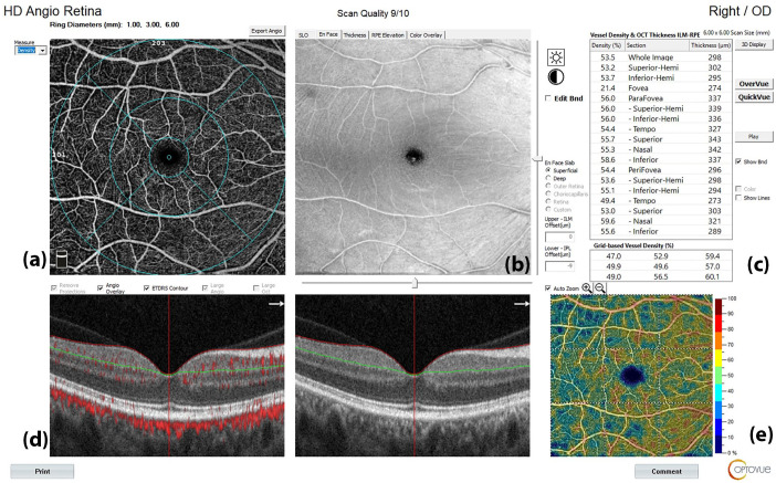
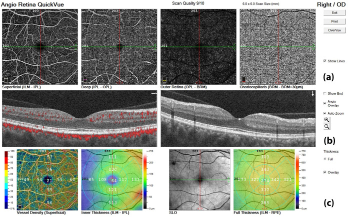
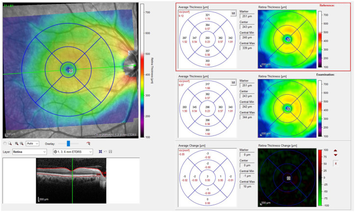
Methods: Patients underwent OCT angiography (OCT-A) to assess retinal vessel density (VD) and spectral-domain OCT (SD-OCT) to evaluate central retinal thickness, macular structure, and peripapillary retinal nerve fiber layer thickness. These parameters were assessed in both groups at T0 and again after 6 months (T1) as part of routine clinical care.
Results: All migraineurs exhibited a significant reduction in disease disability at T1, as assessed by clinical parameters. OCT data analysis revealed that individuals with migraine showed a significant increase in temporal retinal nerve fiber layer (RNFL) thickness and a reduction in nasal RNFL thickness compared to HCs. No differences in retinal circulation were observed between the groups at baseline. At T1, RNFL thickness remained sustained in the superior temporal sector, while the percentage VD of the superficial capillary plexus and radial peripapillary capillary significantly increased in the nasal perifoveal, inferior temporal, and hemi-inferior subregions.
Conclusion: Our study suggests that specific retinal structural changes could precede vascular dysfunction in migraine and can be detected early by combining SD-OCT and OCT-A acquisitions. Short-term treatment with anti-CGRP mAbs may exert neuroprotective effects, potentially preventing permanent ocular damage.
Trial registration: EyeHEAD Study (Trial registration number AIFA July/2024: IT 1735, www.aifa.gov.it/registro-studi-osservazionali).
期刊介绍:
Therapeutic Advances in Neurological Disorders is a peer-reviewed, open access journal delivering the highest quality articles, reviews, and scholarly comment on pioneering efforts and innovative studies across all areas of neurology. The journal has a strong clinical and pharmacological focus and is aimed at clinicians and researchers in neurology, providing a forum in print and online for publishing the highest quality articles in this area.

 求助内容:
求助内容: 应助结果提醒方式:


