Abdulaziz H Almuhanna, Ayman Elnahas, Mohamed K Zabady, Sayed El-Hawari, Mohamed Marzok, Wael Eldeeb, Isam El Jalii, Zakriya Al Mohamad, Arafat Khalaphallah
{"title":"表面健康的小骆驼(骆驼)的瘤胃镜检查:技术描述和瘤胃镜观察。","authors":"Abdulaziz H Almuhanna, Ayman Elnahas, Mohamed K Zabady, Sayed El-Hawari, Mohamed Marzok, Wael Eldeeb, Isam El Jalii, Zakriya Al Mohamad, Arafat Khalaphallah","doi":"10.5455/OVJ.2025.v15.i5.30","DOIUrl":null,"url":null,"abstract":"<p><strong>Background: </strong>Accurate diagnosis of digestive disorders in camels requires validation and optimization of new diagnostic techniques to enable clear visualization of the rumen interior.</p><p><strong>Aim: </strong>The aim of this study was to describe the validity of endoscopy for visualization of the rumen in camels.</p><p><strong>Methods: </strong>Ten apparently healthy camel calves were included in this study. Each camel was appropriately restrained, and the endoscope was inserted through the oro-esophageal route to enable visualization of the rumen.</p><p><strong>Results: </strong>Endoscopy via the oral route allowed visualization of the upper digestive organs and rumen. The ruminal mucosa, glandular parts of the rumen, and ruminal contents were also observed. The technique was performed safely, and no clinical complications were observed.</p><p><strong>Conclusion: </strong>The application of ruminoscopy in camels was shown to be a noninvasive and rapid technique that enabled visual access to the appearance and contents. Such research has the potential to guide clinicians toward better diagnosis of ruminal disorders in the future.</p>","PeriodicalId":19531,"journal":{"name":"Open Veterinary Journal","volume":"15 5","pages":"2122-2126"},"PeriodicalIF":1.0000,"publicationDate":"2025-05-01","publicationTypes":"Journal Article","fieldsOfStudy":null,"isOpenAccess":false,"openAccessPdf":"https://www.ncbi.nlm.nih.gov/pmc/articles/PMC12184463/pdf/","citationCount":"0","resultStr":"{\"title\":\"Ruminoscopy in apparently healthy camel calves (<i>camelus dromedarius</i>): A technique description and ruminoscopic observations.\",\"authors\":\"Abdulaziz H Almuhanna, Ayman Elnahas, Mohamed K Zabady, Sayed El-Hawari, Mohamed Marzok, Wael Eldeeb, Isam El Jalii, Zakriya Al Mohamad, Arafat Khalaphallah\",\"doi\":\"10.5455/OVJ.2025.v15.i5.30\",\"DOIUrl\":null,\"url\":null,\"abstract\":\"<p><strong>Background: </strong>Accurate diagnosis of digestive disorders in camels requires validation and optimization of new diagnostic techniques to enable clear visualization of the rumen interior.</p><p><strong>Aim: </strong>The aim of this study was to describe the validity of endoscopy for visualization of the rumen in camels.</p><p><strong>Methods: </strong>Ten apparently healthy camel calves were included in this study. Each camel was appropriately restrained, and the endoscope was inserted through the oro-esophageal route to enable visualization of the rumen.</p><p><strong>Results: </strong>Endoscopy via the oral route allowed visualization of the upper digestive organs and rumen. The ruminal mucosa, glandular parts of the rumen, and ruminal contents were also observed. The technique was performed safely, and no clinical complications were observed.</p><p><strong>Conclusion: </strong>The application of ruminoscopy in camels was shown to be a noninvasive and rapid technique that enabled visual access to the appearance and contents. Such research has the potential to guide clinicians toward better diagnosis of ruminal disorders in the future.</p>\",\"PeriodicalId\":19531,\"journal\":{\"name\":\"Open Veterinary Journal\",\"volume\":\"15 5\",\"pages\":\"2122-2126\"},\"PeriodicalIF\":1.0000,\"publicationDate\":\"2025-05-01\",\"publicationTypes\":\"Journal Article\",\"fieldsOfStudy\":null,\"isOpenAccess\":false,\"openAccessPdf\":\"https://www.ncbi.nlm.nih.gov/pmc/articles/PMC12184463/pdf/\",\"citationCount\":\"0\",\"resultStr\":null,\"platform\":\"Semanticscholar\",\"paperid\":null,\"PeriodicalName\":\"Open Veterinary Journal\",\"FirstCategoryId\":\"1085\",\"ListUrlMain\":\"https://doi.org/10.5455/OVJ.2025.v15.i5.30\",\"RegionNum\":0,\"RegionCategory\":null,\"ArticlePicture\":[],\"TitleCN\":null,\"AbstractTextCN\":null,\"PMCID\":null,\"EPubDate\":\"2025/5/31 0:00:00\",\"PubModel\":\"Epub\",\"JCR\":\"Q3\",\"JCRName\":\"VETERINARY SCIENCES\",\"Score\":null,\"Total\":0}","platform":"Semanticscholar","paperid":null,"PeriodicalName":"Open Veterinary Journal","FirstCategoryId":"1085","ListUrlMain":"https://doi.org/10.5455/OVJ.2025.v15.i5.30","RegionNum":0,"RegionCategory":null,"ArticlePicture":[],"TitleCN":null,"AbstractTextCN":null,"PMCID":null,"EPubDate":"2025/5/31 0:00:00","PubModel":"Epub","JCR":"Q3","JCRName":"VETERINARY SCIENCES","Score":null,"Total":0}
Ruminoscopy in apparently healthy camel calves (camelus dromedarius): A technique description and ruminoscopic observations.
Background: Accurate diagnosis of digestive disorders in camels requires validation and optimization of new diagnostic techniques to enable clear visualization of the rumen interior.
Aim: The aim of this study was to describe the validity of endoscopy for visualization of the rumen in camels.
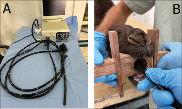
Methods: Ten apparently healthy camel calves were included in this study. Each camel was appropriately restrained, and the endoscope was inserted through the oro-esophageal route to enable visualization of the rumen.
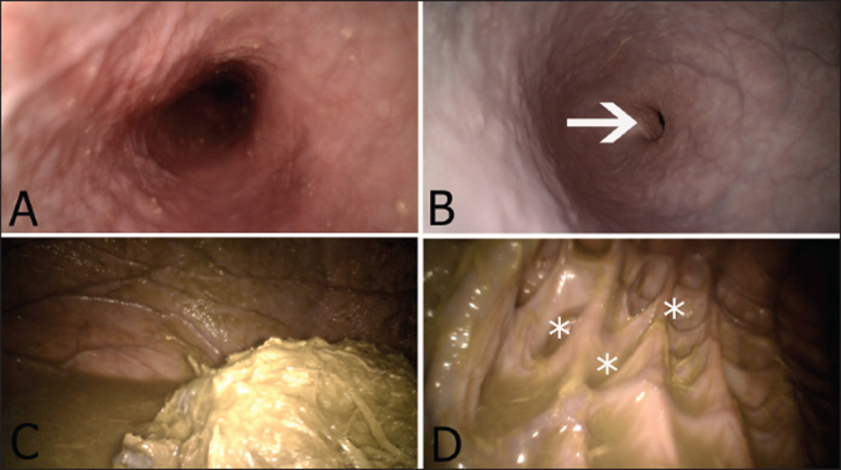
Results: Endoscopy via the oral route allowed visualization of the upper digestive organs and rumen. The ruminal mucosa, glandular parts of the rumen, and ruminal contents were also observed. The technique was performed safely, and no clinical complications were observed.
Conclusion: The application of ruminoscopy in camels was shown to be a noninvasive and rapid technique that enabled visual access to the appearance and contents. Such research has the potential to guide clinicians toward better diagnosis of ruminal disorders in the future.
期刊介绍:
Open Veterinary Journal is a peer-reviewed international open access online and printed journal that publishes high-quality original research articles. reviews, short communications and case reports dedicated to all aspects of veterinary sciences and its related subjects. Research areas include the following: Infectious diseases of zoonotic/food-borne importance, applied biochemistry, parasitology, endocrinology, microbiology, immunology, pathology, pharmacology, physiology, epidemiology, molecular biology, immunogenetics, surgery, ophthalmology, dermatology, oncology and animal reproduction. All papers are peer-reviewed. Moreover, with the presence of well-qualified group of international referees, the process of publication will be done meticulously and to the highest standards.

 求助内容:
求助内容: 应助结果提醒方式:


