Iftikhar Mohammed Abdul Karim, Ramzi Abdulghafoor Al-Agele
{"title":"早孵胚胎(日本鹌鹑和凤尾鸡)和早孵胚胎(赛鸽和凤尾鸟)肢体骨骼发生的组织形态学。","authors":"Iftikhar Mohammed Abdul Karim, Ramzi Abdulghafoor Al-Agele","doi":"10.5455/OVJ.2025.v15.i5.32","DOIUrl":null,"url":null,"abstract":"<p><strong>Background: </strong>Birds are the group of tetrapods that exhibit the greatest diversity in taxonomy and ecology. Limb development is a major focus of developmental and evolutionary biology research.</p><p><strong>Aim: </strong>This study characterized the variances in histomorphometry of skeletogenesis in precocial embryos, such as Japanese quail (Jq) and Cochin chickens (Cc), and altricial embryos, including Racing pigeons (Rp) and Cockatiel birds (Cb).</p><p><strong>Methods: </strong>Six embryos were collected on days 8, 10, 12, 14, 16, and 18 of incubation. Three embryos were prepared and stained with Alcian Blue for chondrification and Alizarin Red for ossified bones. The remaining three embryos were subjected to histological evaluation.</p><p><strong>Results: </strong>Initial signs of ossification appeared in the femur, tibiofibular, and humerus bones of Jq, Rp, and Cb embryos on day 8 and in the bones of Cc on day 10. The statistical study revealed that embryos of different developmental days had considerable variations in average ossified lengths for the humerus of the forelimb, especially Rp, and the femur of the hindlimb, notably in Jq. On day 8, microscopic examination revealed a hypertrophic area with enlarged chondrocytes in the middle and sides of the diaphysis. Osteoblasts significantly augmented the periosteal bone collar around the mid-diaphysis, enhancing its thickness toward the diaphysis center; by day 16, the primary woven bone had developed.</p><p><strong>Conclusion: </strong>This study highlighted the growth rate of the hind limb in precocial embryos, particularly in Jq, which was higher than that in other embryos. These data serve as essential indicators and indispensable parameters for interpreting and elucidating the data collected in these studies.</p>","PeriodicalId":19531,"journal":{"name":"Open Veterinary Journal","volume":"15 5","pages":"2138-2148"},"PeriodicalIF":1.0000,"publicationDate":"2025-05-01","publicationTypes":"Journal Article","fieldsOfStudy":null,"isOpenAccess":false,"openAccessPdf":"https://www.ncbi.nlm.nih.gov/pmc/articles/PMC12184481/pdf/","citationCount":"0","resultStr":"{\"title\":\"Histomorphometry of limb skeletogenesis in prehatched precocial embryos (Japanese quail and Cochin chicken) and altricial embryos (racing pigeons and cockatiel birds).\",\"authors\":\"Iftikhar Mohammed Abdul Karim, Ramzi Abdulghafoor Al-Agele\",\"doi\":\"10.5455/OVJ.2025.v15.i5.32\",\"DOIUrl\":null,\"url\":null,\"abstract\":\"<p><strong>Background: </strong>Birds are the group of tetrapods that exhibit the greatest diversity in taxonomy and ecology. Limb development is a major focus of developmental and evolutionary biology research.</p><p><strong>Aim: </strong>This study characterized the variances in histomorphometry of skeletogenesis in precocial embryos, such as Japanese quail (Jq) and Cochin chickens (Cc), and altricial embryos, including Racing pigeons (Rp) and Cockatiel birds (Cb).</p><p><strong>Methods: </strong>Six embryos were collected on days 8, 10, 12, 14, 16, and 18 of incubation. Three embryos were prepared and stained with Alcian Blue for chondrification and Alizarin Red for ossified bones. The remaining three embryos were subjected to histological evaluation.</p><p><strong>Results: </strong>Initial signs of ossification appeared in the femur, tibiofibular, and humerus bones of Jq, Rp, and Cb embryos on day 8 and in the bones of Cc on day 10. The statistical study revealed that embryos of different developmental days had considerable variations in average ossified lengths for the humerus of the forelimb, especially Rp, and the femur of the hindlimb, notably in Jq. On day 8, microscopic examination revealed a hypertrophic area with enlarged chondrocytes in the middle and sides of the diaphysis. Osteoblasts significantly augmented the periosteal bone collar around the mid-diaphysis, enhancing its thickness toward the diaphysis center; by day 16, the primary woven bone had developed.</p><p><strong>Conclusion: </strong>This study highlighted the growth rate of the hind limb in precocial embryos, particularly in Jq, which was higher than that in other embryos. These data serve as essential indicators and indispensable parameters for interpreting and elucidating the data collected in these studies.</p>\",\"PeriodicalId\":19531,\"journal\":{\"name\":\"Open Veterinary Journal\",\"volume\":\"15 5\",\"pages\":\"2138-2148\"},\"PeriodicalIF\":1.0000,\"publicationDate\":\"2025-05-01\",\"publicationTypes\":\"Journal Article\",\"fieldsOfStudy\":null,\"isOpenAccess\":false,\"openAccessPdf\":\"https://www.ncbi.nlm.nih.gov/pmc/articles/PMC12184481/pdf/\",\"citationCount\":\"0\",\"resultStr\":null,\"platform\":\"Semanticscholar\",\"paperid\":null,\"PeriodicalName\":\"Open Veterinary Journal\",\"FirstCategoryId\":\"1085\",\"ListUrlMain\":\"https://doi.org/10.5455/OVJ.2025.v15.i5.32\",\"RegionNum\":0,\"RegionCategory\":null,\"ArticlePicture\":[],\"TitleCN\":null,\"AbstractTextCN\":null,\"PMCID\":null,\"EPubDate\":\"2025/5/31 0:00:00\",\"PubModel\":\"Epub\",\"JCR\":\"Q3\",\"JCRName\":\"VETERINARY SCIENCES\",\"Score\":null,\"Total\":0}","platform":"Semanticscholar","paperid":null,"PeriodicalName":"Open Veterinary Journal","FirstCategoryId":"1085","ListUrlMain":"https://doi.org/10.5455/OVJ.2025.v15.i5.32","RegionNum":0,"RegionCategory":null,"ArticlePicture":[],"TitleCN":null,"AbstractTextCN":null,"PMCID":null,"EPubDate":"2025/5/31 0:00:00","PubModel":"Epub","JCR":"Q3","JCRName":"VETERINARY SCIENCES","Score":null,"Total":0}
Histomorphometry of limb skeletogenesis in prehatched precocial embryos (Japanese quail and Cochin chicken) and altricial embryos (racing pigeons and cockatiel birds).
Background: Birds are the group of tetrapods that exhibit the greatest diversity in taxonomy and ecology. Limb development is a major focus of developmental and evolutionary biology research.
Aim: This study characterized the variances in histomorphometry of skeletogenesis in precocial embryos, such as Japanese quail (Jq) and Cochin chickens (Cc), and altricial embryos, including Racing pigeons (Rp) and Cockatiel birds (Cb).
Methods: Six embryos were collected on days 8, 10, 12, 14, 16, and 18 of incubation. Three embryos were prepared and stained with Alcian Blue for chondrification and Alizarin Red for ossified bones. The remaining three embryos were subjected to histological evaluation.
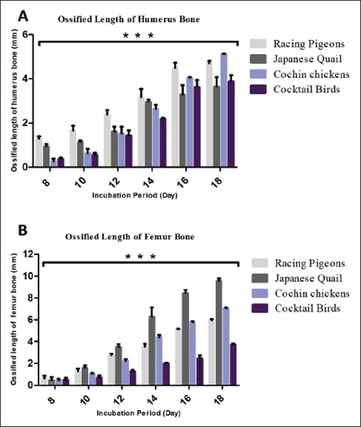
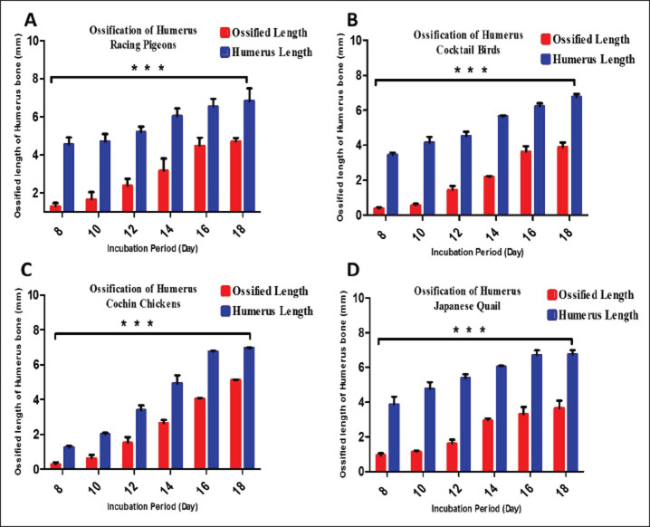
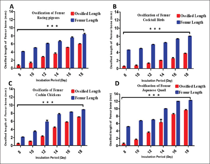
Results: Initial signs of ossification appeared in the femur, tibiofibular, and humerus bones of Jq, Rp, and Cb embryos on day 8 and in the bones of Cc on day 10. The statistical study revealed that embryos of different developmental days had considerable variations in average ossified lengths for the humerus of the forelimb, especially Rp, and the femur of the hindlimb, notably in Jq. On day 8, microscopic examination revealed a hypertrophic area with enlarged chondrocytes in the middle and sides of the diaphysis. Osteoblasts significantly augmented the periosteal bone collar around the mid-diaphysis, enhancing its thickness toward the diaphysis center; by day 16, the primary woven bone had developed.
Conclusion: This study highlighted the growth rate of the hind limb in precocial embryos, particularly in Jq, which was higher than that in other embryos. These data serve as essential indicators and indispensable parameters for interpreting and elucidating the data collected in these studies.
期刊介绍:
Open Veterinary Journal is a peer-reviewed international open access online and printed journal that publishes high-quality original research articles. reviews, short communications and case reports dedicated to all aspects of veterinary sciences and its related subjects. Research areas include the following: Infectious diseases of zoonotic/food-borne importance, applied biochemistry, parasitology, endocrinology, microbiology, immunology, pathology, pharmacology, physiology, epidemiology, molecular biology, immunogenetics, surgery, ophthalmology, dermatology, oncology and animal reproduction. All papers are peer-reviewed. Moreover, with the presence of well-qualified group of international referees, the process of publication will be done meticulously and to the highest standards.

 求助内容:
求助内容: 应助结果提醒方式:


