Aren Dermarderosian, Raffi Boghossian, Derek Tai, Javid Sadjadi, Mojtaba Akhtari
{"title":"应用核医学肝-脾扫描评价脾滨海细胞血管瘤1例。","authors":"Aren Dermarderosian, Raffi Boghossian, Derek Tai, Javid Sadjadi, Mojtaba Akhtari","doi":"10.1177/23247096251350571","DOIUrl":null,"url":null,"abstract":"<p><p>Littoral cell angioma (LCA) is a rare primary splenic vascular neoplasm originating from the littoral cells of the reticuloendothelial system. Splenectomy is the accepted mode of definitive diagnosis and treatment. With fewer than 200 reported cases, LCA remains poorly understood. Herein, we provide an enhanced insight into its histology and highlight the role of nuclear imaging in aiding LCA diagnosis. A 63-year-old female with a history of stage II multiple myeloma (MM) and rheumatoid arthritis was incidentally found to have a slowly enlarging splenic mass over a 6-year period. Given her candidacy for autologous hematopoietic stem cell transplantation for MM, further evaluation of the splenic lesion was pursued using nuclear medicine (NM) liver-spleen scan, which revealed a photopenic region consistent with a benign hemorrhagic mass. Subsequent splenectomy and histopathological analysis confirmed the diagnosis of LCA, with immunohistochemistry demonstrating CD68+ and CD31+ expression, highlighting LCA's unique dual histiocytic and endothelial character. This case highlights the diagnostic challenge posed by LCA due to its nonspecific clinical presentation and imaging findings. While splenectomy remains the gold standard for diagnosis, our findings suggest that NM liver-spleen scan imaging may aid in differentiating LCA from malignant splenic masses preoperatively. Furthermore, this case reinforces the association between LCA and hematologic malignancies, supporting the hypothesis that immune dysregulation may play a role in its pathogenesis. This underscores the importance of considering LCA in the differential diagnosis of splenic masses, particularly in cases involving a history of malignancy and/or immune system abnormalities.</p>","PeriodicalId":16198,"journal":{"name":"Journal of investigative medicine high impact case reports","volume":"13 ","pages":"23247096251350571"},"PeriodicalIF":0.8000,"publicationDate":"2025-01-01","publicationTypes":"Journal Article","fieldsOfStudy":null,"isOpenAccess":false,"openAccessPdf":"https://www.ncbi.nlm.nih.gov/pmc/articles/PMC12198526/pdf/","citationCount":"0","resultStr":"{\"title\":\"Application of Nuclear Medicine Liver-Spleen Scan for Evaluation of Littoral Cell Angioma of the Spleen: A Case Report.\",\"authors\":\"Aren Dermarderosian, Raffi Boghossian, Derek Tai, Javid Sadjadi, Mojtaba Akhtari\",\"doi\":\"10.1177/23247096251350571\",\"DOIUrl\":null,\"url\":null,\"abstract\":\"<p><p>Littoral cell angioma (LCA) is a rare primary splenic vascular neoplasm originating from the littoral cells of the reticuloendothelial system. Splenectomy is the accepted mode of definitive diagnosis and treatment. With fewer than 200 reported cases, LCA remains poorly understood. Herein, we provide an enhanced insight into its histology and highlight the role of nuclear imaging in aiding LCA diagnosis. A 63-year-old female with a history of stage II multiple myeloma (MM) and rheumatoid arthritis was incidentally found to have a slowly enlarging splenic mass over a 6-year period. Given her candidacy for autologous hematopoietic stem cell transplantation for MM, further evaluation of the splenic lesion was pursued using nuclear medicine (NM) liver-spleen scan, which revealed a photopenic region consistent with a benign hemorrhagic mass. Subsequent splenectomy and histopathological analysis confirmed the diagnosis of LCA, with immunohistochemistry demonstrating CD68+ and CD31+ expression, highlighting LCA's unique dual histiocytic and endothelial character. This case highlights the diagnostic challenge posed by LCA due to its nonspecific clinical presentation and imaging findings. While splenectomy remains the gold standard for diagnosis, our findings suggest that NM liver-spleen scan imaging may aid in differentiating LCA from malignant splenic masses preoperatively. Furthermore, this case reinforces the association between LCA and hematologic malignancies, supporting the hypothesis that immune dysregulation may play a role in its pathogenesis. This underscores the importance of considering LCA in the differential diagnosis of splenic masses, particularly in cases involving a history of malignancy and/or immune system abnormalities.</p>\",\"PeriodicalId\":16198,\"journal\":{\"name\":\"Journal of investigative medicine high impact case reports\",\"volume\":\"13 \",\"pages\":\"23247096251350571\"},\"PeriodicalIF\":0.8000,\"publicationDate\":\"2025-01-01\",\"publicationTypes\":\"Journal Article\",\"fieldsOfStudy\":null,\"isOpenAccess\":false,\"openAccessPdf\":\"https://www.ncbi.nlm.nih.gov/pmc/articles/PMC12198526/pdf/\",\"citationCount\":\"0\",\"resultStr\":null,\"platform\":\"Semanticscholar\",\"paperid\":null,\"PeriodicalName\":\"Journal of investigative medicine high impact case reports\",\"FirstCategoryId\":\"1085\",\"ListUrlMain\":\"https://doi.org/10.1177/23247096251350571\",\"RegionNum\":0,\"RegionCategory\":null,\"ArticlePicture\":[],\"TitleCN\":null,\"AbstractTextCN\":null,\"PMCID\":null,\"EPubDate\":\"2025/6/25 0:00:00\",\"PubModel\":\"Epub\",\"JCR\":\"Q3\",\"JCRName\":\"MEDICINE, GENERAL & INTERNAL\",\"Score\":null,\"Total\":0}","platform":"Semanticscholar","paperid":null,"PeriodicalName":"Journal of investigative medicine high impact case reports","FirstCategoryId":"1085","ListUrlMain":"https://doi.org/10.1177/23247096251350571","RegionNum":0,"RegionCategory":null,"ArticlePicture":[],"TitleCN":null,"AbstractTextCN":null,"PMCID":null,"EPubDate":"2025/6/25 0:00:00","PubModel":"Epub","JCR":"Q3","JCRName":"MEDICINE, GENERAL & INTERNAL","Score":null,"Total":0}
Application of Nuclear Medicine Liver-Spleen Scan for Evaluation of Littoral Cell Angioma of the Spleen: A Case Report.
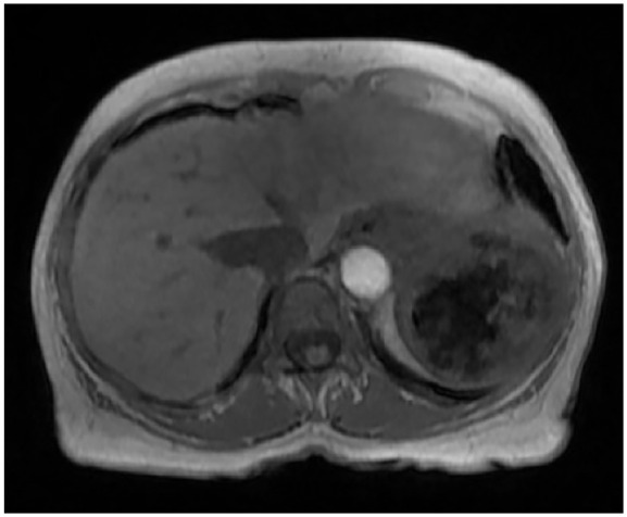
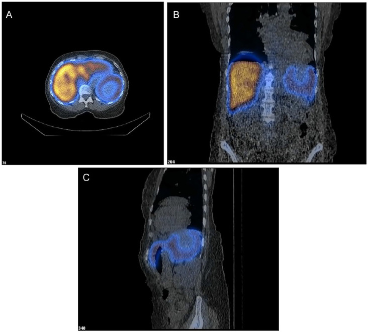
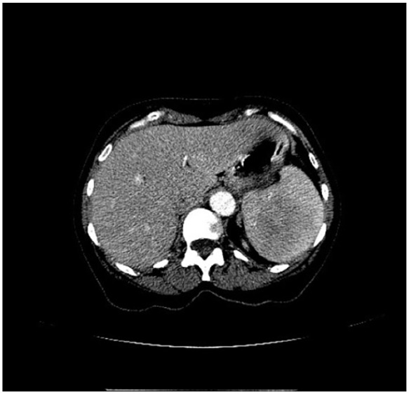
Littoral cell angioma (LCA) is a rare primary splenic vascular neoplasm originating from the littoral cells of the reticuloendothelial system. Splenectomy is the accepted mode of definitive diagnosis and treatment. With fewer than 200 reported cases, LCA remains poorly understood. Herein, we provide an enhanced insight into its histology and highlight the role of nuclear imaging in aiding LCA diagnosis. A 63-year-old female with a history of stage II multiple myeloma (MM) and rheumatoid arthritis was incidentally found to have a slowly enlarging splenic mass over a 6-year period. Given her candidacy for autologous hematopoietic stem cell transplantation for MM, further evaluation of the splenic lesion was pursued using nuclear medicine (NM) liver-spleen scan, which revealed a photopenic region consistent with a benign hemorrhagic mass. Subsequent splenectomy and histopathological analysis confirmed the diagnosis of LCA, with immunohistochemistry demonstrating CD68+ and CD31+ expression, highlighting LCA's unique dual histiocytic and endothelial character. This case highlights the diagnostic challenge posed by LCA due to its nonspecific clinical presentation and imaging findings. While splenectomy remains the gold standard for diagnosis, our findings suggest that NM liver-spleen scan imaging may aid in differentiating LCA from malignant splenic masses preoperatively. Furthermore, this case reinforces the association between LCA and hematologic malignancies, supporting the hypothesis that immune dysregulation may play a role in its pathogenesis. This underscores the importance of considering LCA in the differential diagnosis of splenic masses, particularly in cases involving a history of malignancy and/or immune system abnormalities.
期刊介绍:
The AFMR is committed to enhancing the training and career development of our members and to furthering its mission to facilitate the conduct of research to improve medical care. Case reports represent an important avenue for trainees (interns, residents, and fellows) and early-stage faculty to demonstrate productive, scholarly activity.

 求助内容:
求助内容: 应助结果提醒方式:


