{"title":"非幽门螺杆菌感染引起的胃溃疡。","authors":"Bong Eun Lee","doi":"10.7704/kjhugr.2024.0078","DOIUrl":null,"url":null,"abstract":"<p><p>Gastric ulcers are characterized by mucosal damage extending into the submucosa or deeper, with the most common causes being Helicobacter pylori infection and nonsteroidal anti-inflammatory drug use. However, various infectious pathogens, such as pyogenic bacteria, Treponema pallidum, Mycobacterium tuberculosis, viruses, fungi, and parasites, can also cause gastric ulcers. Non-H. pylori infectious gastric ulcers are uncommon and often present with nonspecific symptoms, making their diagnosis challenging. A differential diagnosis requires a comprehensive understanding of the underlying diseases and familiarity with their characteristic endoscopic features. For instance, acute phlegmonous gastritis requires a prompt diagnosis based on typical clinical symptoms and abdominal computed tomography findings, followed by empiric antibiotic therapy. Infections such as gastric syphilis, gastric tuberculosis, cytomegalovirus (CMV) gastritis, and gastric candidiasis necessitate pathogen identification through tissue diagnoses. When this is challenging, the clinical history, endoscopic findings, and serological tests should be integrated to ensure an accurate diagnosis and management. Unlike gastric syphilis and tuberculosis, CMV gastritis and gastric candidiasis often occur secondary to preexisting gastric ulcers; therefore, conventional anti-ulcer therapy is sufficient for immunocompetent patients with mild symptoms. However, antiviral or antifungal agents should be administered to immunocompromised patients and to those with systemic symptoms related to the infection. Similarly, understanding the characteristic history and symptoms of gastric anisakidosis is crucial for an accurate diagnosis, and prompt endoscopic examination is essential to identify and remove the larvae. Clinicians should consider the possibility of infectious gastric ulcers in patients with atypical ulcerative lesions or ulcers that are unresponsive to conventional therapies. Accurate diagnoses and timely treatments are essential for improving patient outcomes.</p>","PeriodicalId":520887,"journal":{"name":"The Korean journal of helicobacter and upper gastrointestinal research","volume":"25 1","pages":"23-33"},"PeriodicalIF":0.0000,"publicationDate":"2025-03-01","publicationTypes":"Journal Article","fieldsOfStudy":null,"isOpenAccess":false,"openAccessPdf":"https://www.ncbi.nlm.nih.gov/pmc/articles/PMC12173582/pdf/","citationCount":"0","resultStr":"{\"title\":\"Gastric Ulcers Caused by Non-Helicobacter pylori Infections.\",\"authors\":\"Bong Eun Lee\",\"doi\":\"10.7704/kjhugr.2024.0078\",\"DOIUrl\":null,\"url\":null,\"abstract\":\"<p><p>Gastric ulcers are characterized by mucosal damage extending into the submucosa or deeper, with the most common causes being Helicobacter pylori infection and nonsteroidal anti-inflammatory drug use. However, various infectious pathogens, such as pyogenic bacteria, Treponema pallidum, Mycobacterium tuberculosis, viruses, fungi, and parasites, can also cause gastric ulcers. Non-H. pylori infectious gastric ulcers are uncommon and often present with nonspecific symptoms, making their diagnosis challenging. A differential diagnosis requires a comprehensive understanding of the underlying diseases and familiarity with their characteristic endoscopic features. For instance, acute phlegmonous gastritis requires a prompt diagnosis based on typical clinical symptoms and abdominal computed tomography findings, followed by empiric antibiotic therapy. Infections such as gastric syphilis, gastric tuberculosis, cytomegalovirus (CMV) gastritis, and gastric candidiasis necessitate pathogen identification through tissue diagnoses. When this is challenging, the clinical history, endoscopic findings, and serological tests should be integrated to ensure an accurate diagnosis and management. Unlike gastric syphilis and tuberculosis, CMV gastritis and gastric candidiasis often occur secondary to preexisting gastric ulcers; therefore, conventional anti-ulcer therapy is sufficient for immunocompetent patients with mild symptoms. However, antiviral or antifungal agents should be administered to immunocompromised patients and to those with systemic symptoms related to the infection. Similarly, understanding the characteristic history and symptoms of gastric anisakidosis is crucial for an accurate diagnosis, and prompt endoscopic examination is essential to identify and remove the larvae. Clinicians should consider the possibility of infectious gastric ulcers in patients with atypical ulcerative lesions or ulcers that are unresponsive to conventional therapies. Accurate diagnoses and timely treatments are essential for improving patient outcomes.</p>\",\"PeriodicalId\":520887,\"journal\":{\"name\":\"The Korean journal of helicobacter and upper gastrointestinal research\",\"volume\":\"25 1\",\"pages\":\"23-33\"},\"PeriodicalIF\":0.0000,\"publicationDate\":\"2025-03-01\",\"publicationTypes\":\"Journal Article\",\"fieldsOfStudy\":null,\"isOpenAccess\":false,\"openAccessPdf\":\"https://www.ncbi.nlm.nih.gov/pmc/articles/PMC12173582/pdf/\",\"citationCount\":\"0\",\"resultStr\":null,\"platform\":\"Semanticscholar\",\"paperid\":null,\"PeriodicalName\":\"The Korean journal of helicobacter and upper gastrointestinal research\",\"FirstCategoryId\":\"1085\",\"ListUrlMain\":\"https://doi.org/10.7704/kjhugr.2024.0078\",\"RegionNum\":0,\"RegionCategory\":null,\"ArticlePicture\":[],\"TitleCN\":null,\"AbstractTextCN\":null,\"PMCID\":null,\"EPubDate\":\"2025/3/7 0:00:00\",\"PubModel\":\"Epub\",\"JCR\":\"\",\"JCRName\":\"\",\"Score\":null,\"Total\":0}","platform":"Semanticscholar","paperid":null,"PeriodicalName":"The Korean journal of helicobacter and upper gastrointestinal research","FirstCategoryId":"1085","ListUrlMain":"https://doi.org/10.7704/kjhugr.2024.0078","RegionNum":0,"RegionCategory":null,"ArticlePicture":[],"TitleCN":null,"AbstractTextCN":null,"PMCID":null,"EPubDate":"2025/3/7 0:00:00","PubModel":"Epub","JCR":"","JCRName":"","Score":null,"Total":0}
Gastric Ulcers Caused by Non-Helicobacter pylori Infections.
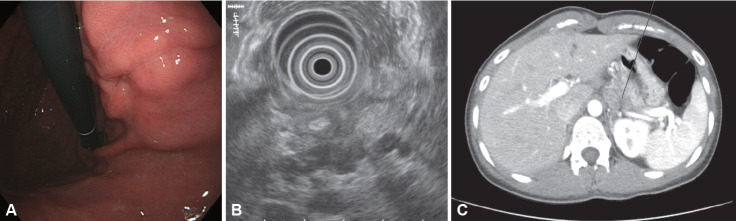
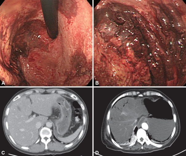
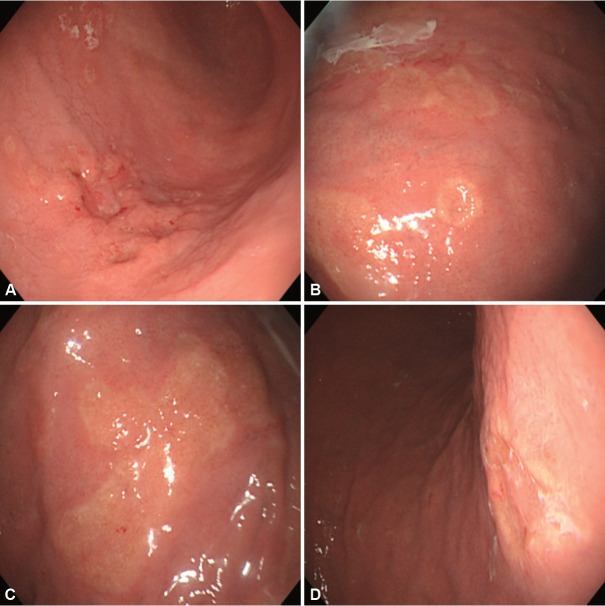
Gastric ulcers are characterized by mucosal damage extending into the submucosa or deeper, with the most common causes being Helicobacter pylori infection and nonsteroidal anti-inflammatory drug use. However, various infectious pathogens, such as pyogenic bacteria, Treponema pallidum, Mycobacterium tuberculosis, viruses, fungi, and parasites, can also cause gastric ulcers. Non-H. pylori infectious gastric ulcers are uncommon and often present with nonspecific symptoms, making their diagnosis challenging. A differential diagnosis requires a comprehensive understanding of the underlying diseases and familiarity with their characteristic endoscopic features. For instance, acute phlegmonous gastritis requires a prompt diagnosis based on typical clinical symptoms and abdominal computed tomography findings, followed by empiric antibiotic therapy. Infections such as gastric syphilis, gastric tuberculosis, cytomegalovirus (CMV) gastritis, and gastric candidiasis necessitate pathogen identification through tissue diagnoses. When this is challenging, the clinical history, endoscopic findings, and serological tests should be integrated to ensure an accurate diagnosis and management. Unlike gastric syphilis and tuberculosis, CMV gastritis and gastric candidiasis often occur secondary to preexisting gastric ulcers; therefore, conventional anti-ulcer therapy is sufficient for immunocompetent patients with mild symptoms. However, antiviral or antifungal agents should be administered to immunocompromised patients and to those with systemic symptoms related to the infection. Similarly, understanding the characteristic history and symptoms of gastric anisakidosis is crucial for an accurate diagnosis, and prompt endoscopic examination is essential to identify and remove the larvae. Clinicians should consider the possibility of infectious gastric ulcers in patients with atypical ulcerative lesions or ulcers that are unresponsive to conventional therapies. Accurate diagnoses and timely treatments are essential for improving patient outcomes.

 求助内容:
求助内容: 应助结果提醒方式:


