Ioannis Loufopoulos, Clarence Zwengunde, Soumendra Datta
{"title":"双肾收集系统结石:一个具有挑战性的管理技术。","authors":"Ioannis Loufopoulos, Clarence Zwengunde, Soumendra Datta","doi":"10.1155/criu/1543018","DOIUrl":null,"url":null,"abstract":"<p><p>Duplex renal collecting system is a relatively common congenital abnormality affecting equally both pelvicalyceal systems. Although usually it is an incidental finding, it can cause significant problems to the patients such as recurrent urinary tract infections, hydronephrosis, and lithiasis. In this study, we describe an interesting surgical management option for a patient with upper moiety hydronephrosis and lithiasis of the aberrant ureter, achieving resolution of the hydronephrosis and complete removal of the calculus. A 49-year-old female patient presented with symptoms of left colicky pain. During the initial investigation, a left-sided duplex renal collecting system with severely hydronephrotic upper pole moiety and grossly dilated tortuous ureter with distal calculus and ectopic insertion to urinary bladder was identified. On cystoscopy, the upper moiety ureteric opening was identified distally to the urethral sphincter. Under ultrasound guidance, endoscopic transvesical resection was performed distal to the stented lower moiety ureteric orifice, resulting in the identification of the stone and extraction. Short- and long-term follow-up demonstrated no recurrence of the stone and significant resolution of the hydronephrosis. In conclusion, in this case report, we describe an unusual anatomical variation of the upper moiety outflow, and we introduce a new technique of intravesical ultrasound-guided removal of an obstructive calculus.</p>","PeriodicalId":30323,"journal":{"name":"Case Reports in Urology","volume":"2025 ","pages":"1543018"},"PeriodicalIF":0.0000,"publicationDate":"2025-06-12","publicationTypes":"Journal Article","fieldsOfStudy":null,"isOpenAccess":false,"openAccessPdf":"https://www.ncbi.nlm.nih.gov/pmc/articles/PMC12178762/pdf/","citationCount":"0","resultStr":"{\"title\":\"Duplex Renal Collecting System Lithiasis: A Case of a Challenging Management Technique.\",\"authors\":\"Ioannis Loufopoulos, Clarence Zwengunde, Soumendra Datta\",\"doi\":\"10.1155/criu/1543018\",\"DOIUrl\":null,\"url\":null,\"abstract\":\"<p><p>Duplex renal collecting system is a relatively common congenital abnormality affecting equally both pelvicalyceal systems. Although usually it is an incidental finding, it can cause significant problems to the patients such as recurrent urinary tract infections, hydronephrosis, and lithiasis. In this study, we describe an interesting surgical management option for a patient with upper moiety hydronephrosis and lithiasis of the aberrant ureter, achieving resolution of the hydronephrosis and complete removal of the calculus. A 49-year-old female patient presented with symptoms of left colicky pain. During the initial investigation, a left-sided duplex renal collecting system with severely hydronephrotic upper pole moiety and grossly dilated tortuous ureter with distal calculus and ectopic insertion to urinary bladder was identified. On cystoscopy, the upper moiety ureteric opening was identified distally to the urethral sphincter. Under ultrasound guidance, endoscopic transvesical resection was performed distal to the stented lower moiety ureteric orifice, resulting in the identification of the stone and extraction. Short- and long-term follow-up demonstrated no recurrence of the stone and significant resolution of the hydronephrosis. In conclusion, in this case report, we describe an unusual anatomical variation of the upper moiety outflow, and we introduce a new technique of intravesical ultrasound-guided removal of an obstructive calculus.</p>\",\"PeriodicalId\":30323,\"journal\":{\"name\":\"Case Reports in Urology\",\"volume\":\"2025 \",\"pages\":\"1543018\"},\"PeriodicalIF\":0.0000,\"publicationDate\":\"2025-06-12\",\"publicationTypes\":\"Journal Article\",\"fieldsOfStudy\":null,\"isOpenAccess\":false,\"openAccessPdf\":\"https://www.ncbi.nlm.nih.gov/pmc/articles/PMC12178762/pdf/\",\"citationCount\":\"0\",\"resultStr\":null,\"platform\":\"Semanticscholar\",\"paperid\":null,\"PeriodicalName\":\"Case Reports in Urology\",\"FirstCategoryId\":\"1085\",\"ListUrlMain\":\"https://doi.org/10.1155/criu/1543018\",\"RegionNum\":0,\"RegionCategory\":null,\"ArticlePicture\":[],\"TitleCN\":null,\"AbstractTextCN\":null,\"PMCID\":null,\"EPubDate\":\"2025/1/1 0:00:00\",\"PubModel\":\"eCollection\",\"JCR\":\"\",\"JCRName\":\"\",\"Score\":null,\"Total\":0}","platform":"Semanticscholar","paperid":null,"PeriodicalName":"Case Reports in Urology","FirstCategoryId":"1085","ListUrlMain":"https://doi.org/10.1155/criu/1543018","RegionNum":0,"RegionCategory":null,"ArticlePicture":[],"TitleCN":null,"AbstractTextCN":null,"PMCID":null,"EPubDate":"2025/1/1 0:00:00","PubModel":"eCollection","JCR":"","JCRName":"","Score":null,"Total":0}
Duplex Renal Collecting System Lithiasis: A Case of a Challenging Management Technique.
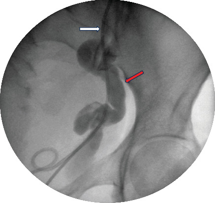
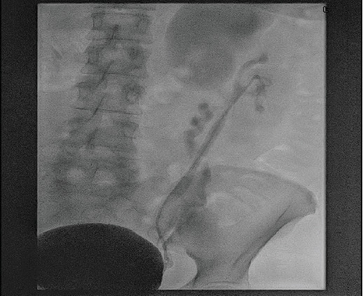
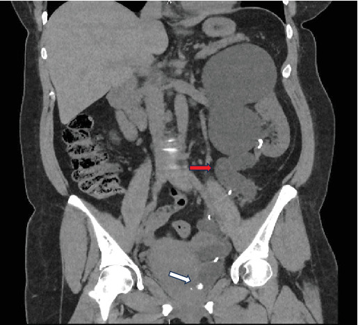
Duplex renal collecting system is a relatively common congenital abnormality affecting equally both pelvicalyceal systems. Although usually it is an incidental finding, it can cause significant problems to the patients such as recurrent urinary tract infections, hydronephrosis, and lithiasis. In this study, we describe an interesting surgical management option for a patient with upper moiety hydronephrosis and lithiasis of the aberrant ureter, achieving resolution of the hydronephrosis and complete removal of the calculus. A 49-year-old female patient presented with symptoms of left colicky pain. During the initial investigation, a left-sided duplex renal collecting system with severely hydronephrotic upper pole moiety and grossly dilated tortuous ureter with distal calculus and ectopic insertion to urinary bladder was identified. On cystoscopy, the upper moiety ureteric opening was identified distally to the urethral sphincter. Under ultrasound guidance, endoscopic transvesical resection was performed distal to the stented lower moiety ureteric orifice, resulting in the identification of the stone and extraction. Short- and long-term follow-up demonstrated no recurrence of the stone and significant resolution of the hydronephrosis. In conclusion, in this case report, we describe an unusual anatomical variation of the upper moiety outflow, and we introduce a new technique of intravesical ultrasound-guided removal of an obstructive calculus.

 求助内容:
求助内容: 应助结果提醒方式:


