Heli Tuomainen, Mazen Sudah, Sarianna Joukainen, Vesa Kärjä, Amro Masarwah, Otto Jokelainen, Hidemi Okuma
{"title":"乳房x光检查发现的与恶性乳腺肿瘤相关的针状体经常含有额外的肿瘤灶。","authors":"Heli Tuomainen, Mazen Sudah, Sarianna Joukainen, Vesa Kärjä, Amro Masarwah, Otto Jokelainen, Hidemi Okuma","doi":"10.2478/raon-2025-0041","DOIUrl":null,"url":null,"abstract":"<p><strong>Background: </strong>On imaging, malignant breast masses are commonly associated with spicules. To the best of our knowledge, the clinical significance of such spiculae has not been previously studied, and no surgical guidelines are available for the management of mammographically detected spiculations.</p><p><strong>Patients and methods: </strong>Between April 2018 and December 2019, all consecutive breast-conserving surgery -patients with invasive malignant lesions, who required intraoperative radiological breast specimen assessment with tomosynthesis, were retrospectively included in this analysis. The tumors were classified into two groups: those with spiculated margins as the dominant feature, and those with other distinct morphological characteristics. Spicule visualization, length, and distribution were evaluated in both groups using pre- and intraoperative imaging and compared with the histopathological features of the spicules.</p><p><strong>Results: </strong>In total, 162 invasive lesions were evaluated. The presence of spicule-associated additional tumor foci was a common finding; 67.6% of the spiculated tumors and 48.9% of the other tumors had additional foci. Most additional tumor foci were within 1 cm of the tumor edge. The mean pathologically measured distance from the main tumor margin to the spicule-associated additional tumor foci was 4.3 ± 2.8 mm. Compared to the maximum spicule length determined from intraoperative images (9.5 ± 5.1 mm), the distance of actual tumor infiltration was much shorter, and a very weak correlation was observed.</p><p><strong>Conclusions: </strong>Breast tumor spicules harbor additional tumor foci, which may lead to margin positivity and potential reoperation. Additional research is necessary to determine the actual tumor burden and clinical significance of spicules.</p>","PeriodicalId":21034,"journal":{"name":"Radiology and Oncology","volume":"59 2","pages":"168-175"},"PeriodicalIF":2.2000,"publicationDate":"2025-06-21","publicationTypes":"Journal Article","fieldsOfStudy":null,"isOpenAccess":false,"openAccessPdf":"https://www.ncbi.nlm.nih.gov/pmc/articles/PMC12182920/pdf/","citationCount":"0","resultStr":"{\"title\":\"Mammographically detected spicules associated with malignant breast tumors frequently harbor additional tumor foci.\",\"authors\":\"Heli Tuomainen, Mazen Sudah, Sarianna Joukainen, Vesa Kärjä, Amro Masarwah, Otto Jokelainen, Hidemi Okuma\",\"doi\":\"10.2478/raon-2025-0041\",\"DOIUrl\":null,\"url\":null,\"abstract\":\"<p><strong>Background: </strong>On imaging, malignant breast masses are commonly associated with spicules. To the best of our knowledge, the clinical significance of such spiculae has not been previously studied, and no surgical guidelines are available for the management of mammographically detected spiculations.</p><p><strong>Patients and methods: </strong>Between April 2018 and December 2019, all consecutive breast-conserving surgery -patients with invasive malignant lesions, who required intraoperative radiological breast specimen assessment with tomosynthesis, were retrospectively included in this analysis. The tumors were classified into two groups: those with spiculated margins as the dominant feature, and those with other distinct morphological characteristics. Spicule visualization, length, and distribution were evaluated in both groups using pre- and intraoperative imaging and compared with the histopathological features of the spicules.</p><p><strong>Results: </strong>In total, 162 invasive lesions were evaluated. The presence of spicule-associated additional tumor foci was a common finding; 67.6% of the spiculated tumors and 48.9% of the other tumors had additional foci. Most additional tumor foci were within 1 cm of the tumor edge. The mean pathologically measured distance from the main tumor margin to the spicule-associated additional tumor foci was 4.3 ± 2.8 mm. Compared to the maximum spicule length determined from intraoperative images (9.5 ± 5.1 mm), the distance of actual tumor infiltration was much shorter, and a very weak correlation was observed.</p><p><strong>Conclusions: </strong>Breast tumor spicules harbor additional tumor foci, which may lead to margin positivity and potential reoperation. Additional research is necessary to determine the actual tumor burden and clinical significance of spicules.</p>\",\"PeriodicalId\":21034,\"journal\":{\"name\":\"Radiology and Oncology\",\"volume\":\"59 2\",\"pages\":\"168-175\"},\"PeriodicalIF\":2.2000,\"publicationDate\":\"2025-06-21\",\"publicationTypes\":\"Journal Article\",\"fieldsOfStudy\":null,\"isOpenAccess\":false,\"openAccessPdf\":\"https://www.ncbi.nlm.nih.gov/pmc/articles/PMC12182920/pdf/\",\"citationCount\":\"0\",\"resultStr\":null,\"platform\":\"Semanticscholar\",\"paperid\":null,\"PeriodicalName\":\"Radiology and Oncology\",\"FirstCategoryId\":\"3\",\"ListUrlMain\":\"https://doi.org/10.2478/raon-2025-0041\",\"RegionNum\":4,\"RegionCategory\":\"医学\",\"ArticlePicture\":[],\"TitleCN\":null,\"AbstractTextCN\":null,\"PMCID\":null,\"EPubDate\":\"2025/6/1 0:00:00\",\"PubModel\":\"eCollection\",\"JCR\":\"Q3\",\"JCRName\":\"ONCOLOGY\",\"Score\":null,\"Total\":0}","platform":"Semanticscholar","paperid":null,"PeriodicalName":"Radiology and Oncology","FirstCategoryId":"3","ListUrlMain":"https://doi.org/10.2478/raon-2025-0041","RegionNum":4,"RegionCategory":"医学","ArticlePicture":[],"TitleCN":null,"AbstractTextCN":null,"PMCID":null,"EPubDate":"2025/6/1 0:00:00","PubModel":"eCollection","JCR":"Q3","JCRName":"ONCOLOGY","Score":null,"Total":0}
Mammographically detected spicules associated with malignant breast tumors frequently harbor additional tumor foci.
Background: On imaging, malignant breast masses are commonly associated with spicules. To the best of our knowledge, the clinical significance of such spiculae has not been previously studied, and no surgical guidelines are available for the management of mammographically detected spiculations.
Patients and methods: Between April 2018 and December 2019, all consecutive breast-conserving surgery -patients with invasive malignant lesions, who required intraoperative radiological breast specimen assessment with tomosynthesis, were retrospectively included in this analysis. The tumors were classified into two groups: those with spiculated margins as the dominant feature, and those with other distinct morphological characteristics. Spicule visualization, length, and distribution were evaluated in both groups using pre- and intraoperative imaging and compared with the histopathological features of the spicules.
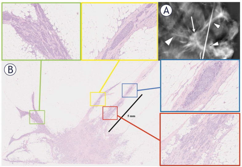
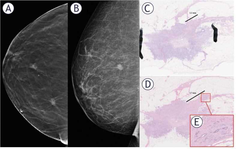
Results: In total, 162 invasive lesions were evaluated. The presence of spicule-associated additional tumor foci was a common finding; 67.6% of the spiculated tumors and 48.9% of the other tumors had additional foci. Most additional tumor foci were within 1 cm of the tumor edge. The mean pathologically measured distance from the main tumor margin to the spicule-associated additional tumor foci was 4.3 ± 2.8 mm. Compared to the maximum spicule length determined from intraoperative images (9.5 ± 5.1 mm), the distance of actual tumor infiltration was much shorter, and a very weak correlation was observed.
Conclusions: Breast tumor spicules harbor additional tumor foci, which may lead to margin positivity and potential reoperation. Additional research is necessary to determine the actual tumor burden and clinical significance of spicules.
期刊介绍:
Radiology and Oncology is a multidisciplinary journal devoted to the publishing original and high quality scientific papers and review articles, pertinent to diagnostic and interventional radiology, computerized tomography, magnetic resonance, ultrasound, nuclear medicine, radiotherapy, clinical and experimental oncology, radiobiology, medical physics and radiation protection. Therefore, the scope of the journal is to cover beside radiology the diagnostic and therapeutic aspects in oncology, which distinguishes it from other journals in the field.

 求助内容:
求助内容: 应助结果提醒方式:


