Ahmad Al-Bitar, Bishr A Al-Abdulrazzak, Ruba Alahmar
{"title":"鼻窦腺样囊性癌1例报告及文献复习。","authors":"Ahmad Al-Bitar, Bishr A Al-Abdulrazzak, Ruba Alahmar","doi":"10.1159/000546446","DOIUrl":null,"url":null,"abstract":"<p><strong>Introduction: </strong>Adenoid cystic carcinoma (ACC), a rare, aggressive malignancy of secretory epithelia, is often present with nonspecific symptoms, delaying diagnosis.</p><p><strong>Case presentation: </strong>A 67-year-old male reported 10 months of nasal obstruction, mouth breathing, and sleep disturbances. The initial evaluation for foreign body obstruction revealed septal deviation and low-density sinonasal tissue on CT. MRI identified a 7 × 4.5 × 7 cm heterogeneous lesion invading nasal structures, paranasal sinuses, and nasopharynx with diffusion restriction. Tru-cut biopsy confirmed ACC via cribriform, tubular, and solid basaloid cell patterns, pseudocystic spaces, biphasic ductal-myoepithelial cells, and perineural invasion. Immunohistochemistry (CK7, CD117, p63, S100) supported the diagnosis, with tumor-free margins and no metastases. Multimodal therapy (30 VMAT sessions, 4 cisplatin-vinorelbine cycles) improved symptoms.</p><p><strong>Conclusion: </strong>This case illustrates ACC's diagnostic complexity, requiring advanced imaging and histopathology to exclude mimics. Despite indolent early progression, ACC's neurotropic spread and late-stage detection demand aggressive treatment. While surgery with adjuvant radiotherapy remains standard, the absence of metastases here underscores its variable behavior.</p>","PeriodicalId":9625,"journal":{"name":"Case Reports in Oncology","volume":"18 1","pages":"809-816"},"PeriodicalIF":0.7000,"publicationDate":"2025-05-28","publicationTypes":"Journal Article","fieldsOfStudy":null,"isOpenAccess":false,"openAccessPdf":"https://www.ncbi.nlm.nih.gov/pmc/articles/PMC12180789/pdf/","citationCount":"0","resultStr":"{\"title\":\"Adenoid Cystic Carcinoma of the Sinonasal Cavity: A Case Report and Literature Review.\",\"authors\":\"Ahmad Al-Bitar, Bishr A Al-Abdulrazzak, Ruba Alahmar\",\"doi\":\"10.1159/000546446\",\"DOIUrl\":null,\"url\":null,\"abstract\":\"<p><strong>Introduction: </strong>Adenoid cystic carcinoma (ACC), a rare, aggressive malignancy of secretory epithelia, is often present with nonspecific symptoms, delaying diagnosis.</p><p><strong>Case presentation: </strong>A 67-year-old male reported 10 months of nasal obstruction, mouth breathing, and sleep disturbances. The initial evaluation for foreign body obstruction revealed septal deviation and low-density sinonasal tissue on CT. MRI identified a 7 × 4.5 × 7 cm heterogeneous lesion invading nasal structures, paranasal sinuses, and nasopharynx with diffusion restriction. Tru-cut biopsy confirmed ACC via cribriform, tubular, and solid basaloid cell patterns, pseudocystic spaces, biphasic ductal-myoepithelial cells, and perineural invasion. Immunohistochemistry (CK7, CD117, p63, S100) supported the diagnosis, with tumor-free margins and no metastases. Multimodal therapy (30 VMAT sessions, 4 cisplatin-vinorelbine cycles) improved symptoms.</p><p><strong>Conclusion: </strong>This case illustrates ACC's diagnostic complexity, requiring advanced imaging and histopathology to exclude mimics. Despite indolent early progression, ACC's neurotropic spread and late-stage detection demand aggressive treatment. While surgery with adjuvant radiotherapy remains standard, the absence of metastases here underscores its variable behavior.</p>\",\"PeriodicalId\":9625,\"journal\":{\"name\":\"Case Reports in Oncology\",\"volume\":\"18 1\",\"pages\":\"809-816\"},\"PeriodicalIF\":0.7000,\"publicationDate\":\"2025-05-28\",\"publicationTypes\":\"Journal Article\",\"fieldsOfStudy\":null,\"isOpenAccess\":false,\"openAccessPdf\":\"https://www.ncbi.nlm.nih.gov/pmc/articles/PMC12180789/pdf/\",\"citationCount\":\"0\",\"resultStr\":null,\"platform\":\"Semanticscholar\",\"paperid\":null,\"PeriodicalName\":\"Case Reports in Oncology\",\"FirstCategoryId\":\"1085\",\"ListUrlMain\":\"https://doi.org/10.1159/000546446\",\"RegionNum\":0,\"RegionCategory\":null,\"ArticlePicture\":[],\"TitleCN\":null,\"AbstractTextCN\":null,\"PMCID\":null,\"EPubDate\":\"2025/1/1 0:00:00\",\"PubModel\":\"eCollection\",\"JCR\":\"Q4\",\"JCRName\":\"ONCOLOGY\",\"Score\":null,\"Total\":0}","platform":"Semanticscholar","paperid":null,"PeriodicalName":"Case Reports in Oncology","FirstCategoryId":"1085","ListUrlMain":"https://doi.org/10.1159/000546446","RegionNum":0,"RegionCategory":null,"ArticlePicture":[],"TitleCN":null,"AbstractTextCN":null,"PMCID":null,"EPubDate":"2025/1/1 0:00:00","PubModel":"eCollection","JCR":"Q4","JCRName":"ONCOLOGY","Score":null,"Total":0}
Adenoid Cystic Carcinoma of the Sinonasal Cavity: A Case Report and Literature Review.
Introduction: Adenoid cystic carcinoma (ACC), a rare, aggressive malignancy of secretory epithelia, is often present with nonspecific symptoms, delaying diagnosis.
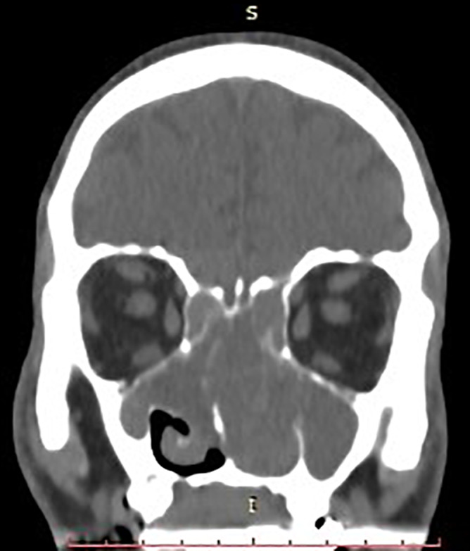
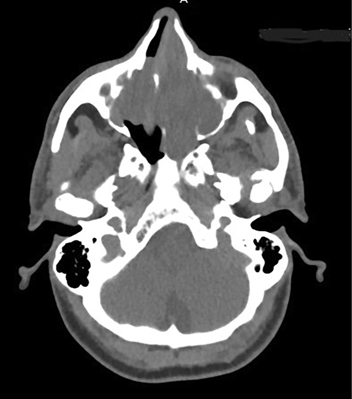
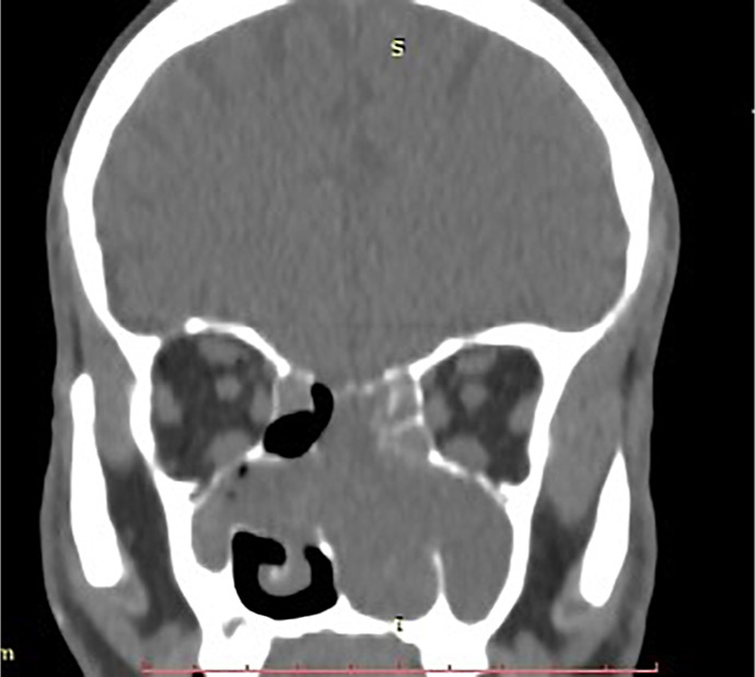
Case presentation: A 67-year-old male reported 10 months of nasal obstruction, mouth breathing, and sleep disturbances. The initial evaluation for foreign body obstruction revealed septal deviation and low-density sinonasal tissue on CT. MRI identified a 7 × 4.5 × 7 cm heterogeneous lesion invading nasal structures, paranasal sinuses, and nasopharynx with diffusion restriction. Tru-cut biopsy confirmed ACC via cribriform, tubular, and solid basaloid cell patterns, pseudocystic spaces, biphasic ductal-myoepithelial cells, and perineural invasion. Immunohistochemistry (CK7, CD117, p63, S100) supported the diagnosis, with tumor-free margins and no metastases. Multimodal therapy (30 VMAT sessions, 4 cisplatin-vinorelbine cycles) improved symptoms.
Conclusion: This case illustrates ACC's diagnostic complexity, requiring advanced imaging and histopathology to exclude mimics. Despite indolent early progression, ACC's neurotropic spread and late-stage detection demand aggressive treatment. While surgery with adjuvant radiotherapy remains standard, the absence of metastases here underscores its variable behavior.

 求助内容:
求助内容: 应助结果提醒方式:


