Suwei Liu, Yali Li, Shuai Tian, Chenyu Jiang, Ming Ni, Ke Xu, Feng Wei, Huishu Yuan
{"title":"CT δ放射组学预测脊柱肿瘤手术中输血和大出血的风险。","authors":"Suwei Liu, Yali Li, Shuai Tian, Chenyu Jiang, Ming Ni, Ke Xu, Feng Wei, Huishu Yuan","doi":"10.1186/s40644-025-00900-1","DOIUrl":null,"url":null,"abstract":"<p><strong>Background: </strong>Intraoperative bleeding is a serious complication of spinal tumor surgery. Preoperative identification of patients at high risk of intraoperative blood transfusion (IBT) and intraoperative massive bleeding (IMB) before spinal tumor resection surgery is difficult but critical for surgical planning and blood management. This study aims to develop and validate delta radiomics prediction models for IBT and IMB in spinal tumor surgery.</p><p><strong>Methods: </strong>Patients diagnosed with spinal tumors who underwent spinal tumor resection surgery were retrospectively recruited. CT, CTE, delta, and clinical models based on CT native phase, CT arterial phase images, and clinical factors were constructed using 10-fold cross-validation and logistic regression (LR), random forest (RF), and support vector machine (SVM) in the training cohort. Receiver operating characteristic (ROC) curves, integrated discrimination improvement (IDI), accuracy, sensitivity, specificity, positive predictive value, and negative predictive value were used to evaluate and compare the diagnostic performance of these models.</p><p><strong>Results: </strong>231 patients were randomly divided into training (n = 161) and test (n = 70) cohorts, comprising 146 IBT and 85 no-IBT patients, 35 IMB and 196 no-IMB patients, respectively. The delta model performed best in predicting IBT and IMB risk, with better predictive ability than the clinical model (IDI = 0.11-0.13 for IBT, and IDI = 0.02-0.08 for IMB, p < 0.05, respectively). Calibration curves indicated that the predicted probabilities of IBT and IMB in the model did not differ significantly from the actual probabilities (p > 0.05).</p><p><strong>Conclusion: </strong>The CT delta model we constructed may be a valuable tool to improve risk stratification before spinal tumor surgery, thus contributing to preoperative planning and improving patient prognosis.</p><p><strong>Trial registration: </strong>Retrospectively registered (M2020435).</p>","PeriodicalId":9548,"journal":{"name":"Cancer Imaging","volume":"25 1","pages":"79"},"PeriodicalIF":3.5000,"publicationDate":"2025-06-22","publicationTypes":"Journal Article","fieldsOfStudy":null,"isOpenAccess":false,"openAccessPdf":"https://www.ncbi.nlm.nih.gov/pmc/articles/PMC12183832/pdf/","citationCount":"0","resultStr":"{\"title\":\"CT delta-radiomics predicts the risks of blood transfusion and massive bleeding during spinal tumor surgery.\",\"authors\":\"Suwei Liu, Yali Li, Shuai Tian, Chenyu Jiang, Ming Ni, Ke Xu, Feng Wei, Huishu Yuan\",\"doi\":\"10.1186/s40644-025-00900-1\",\"DOIUrl\":null,\"url\":null,\"abstract\":\"<p><strong>Background: </strong>Intraoperative bleeding is a serious complication of spinal tumor surgery. Preoperative identification of patients at high risk of intraoperative blood transfusion (IBT) and intraoperative massive bleeding (IMB) before spinal tumor resection surgery is difficult but critical for surgical planning and blood management. This study aims to develop and validate delta radiomics prediction models for IBT and IMB in spinal tumor surgery.</p><p><strong>Methods: </strong>Patients diagnosed with spinal tumors who underwent spinal tumor resection surgery were retrospectively recruited. CT, CTE, delta, and clinical models based on CT native phase, CT arterial phase images, and clinical factors were constructed using 10-fold cross-validation and logistic regression (LR), random forest (RF), and support vector machine (SVM) in the training cohort. Receiver operating characteristic (ROC) curves, integrated discrimination improvement (IDI), accuracy, sensitivity, specificity, positive predictive value, and negative predictive value were used to evaluate and compare the diagnostic performance of these models.</p><p><strong>Results: </strong>231 patients were randomly divided into training (n = 161) and test (n = 70) cohorts, comprising 146 IBT and 85 no-IBT patients, 35 IMB and 196 no-IMB patients, respectively. The delta model performed best in predicting IBT and IMB risk, with better predictive ability than the clinical model (IDI = 0.11-0.13 for IBT, and IDI = 0.02-0.08 for IMB, p < 0.05, respectively). Calibration curves indicated that the predicted probabilities of IBT and IMB in the model did not differ significantly from the actual probabilities (p > 0.05).</p><p><strong>Conclusion: </strong>The CT delta model we constructed may be a valuable tool to improve risk stratification before spinal tumor surgery, thus contributing to preoperative planning and improving patient prognosis.</p><p><strong>Trial registration: </strong>Retrospectively registered (M2020435).</p>\",\"PeriodicalId\":9548,\"journal\":{\"name\":\"Cancer Imaging\",\"volume\":\"25 1\",\"pages\":\"79\"},\"PeriodicalIF\":3.5000,\"publicationDate\":\"2025-06-22\",\"publicationTypes\":\"Journal Article\",\"fieldsOfStudy\":null,\"isOpenAccess\":false,\"openAccessPdf\":\"https://www.ncbi.nlm.nih.gov/pmc/articles/PMC12183832/pdf/\",\"citationCount\":\"0\",\"resultStr\":null,\"platform\":\"Semanticscholar\",\"paperid\":null,\"PeriodicalName\":\"Cancer Imaging\",\"FirstCategoryId\":\"3\",\"ListUrlMain\":\"https://doi.org/10.1186/s40644-025-00900-1\",\"RegionNum\":2,\"RegionCategory\":\"医学\",\"ArticlePicture\":[],\"TitleCN\":null,\"AbstractTextCN\":null,\"PMCID\":null,\"EPubDate\":\"\",\"PubModel\":\"\",\"JCR\":\"Q2\",\"JCRName\":\"ONCOLOGY\",\"Score\":null,\"Total\":0}","platform":"Semanticscholar","paperid":null,"PeriodicalName":"Cancer Imaging","FirstCategoryId":"3","ListUrlMain":"https://doi.org/10.1186/s40644-025-00900-1","RegionNum":2,"RegionCategory":"医学","ArticlePicture":[],"TitleCN":null,"AbstractTextCN":null,"PMCID":null,"EPubDate":"","PubModel":"","JCR":"Q2","JCRName":"ONCOLOGY","Score":null,"Total":0}
CT delta-radiomics predicts the risks of blood transfusion and massive bleeding during spinal tumor surgery.
Background: Intraoperative bleeding is a serious complication of spinal tumor surgery. Preoperative identification of patients at high risk of intraoperative blood transfusion (IBT) and intraoperative massive bleeding (IMB) before spinal tumor resection surgery is difficult but critical for surgical planning and blood management. This study aims to develop and validate delta radiomics prediction models for IBT and IMB in spinal tumor surgery.
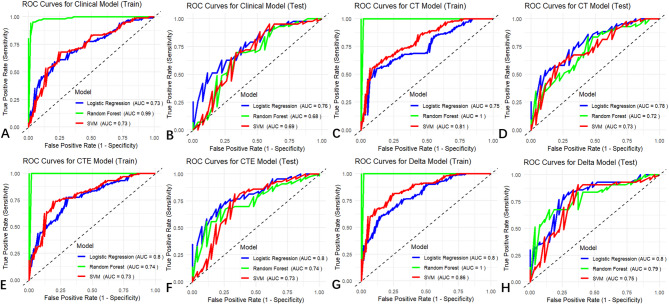
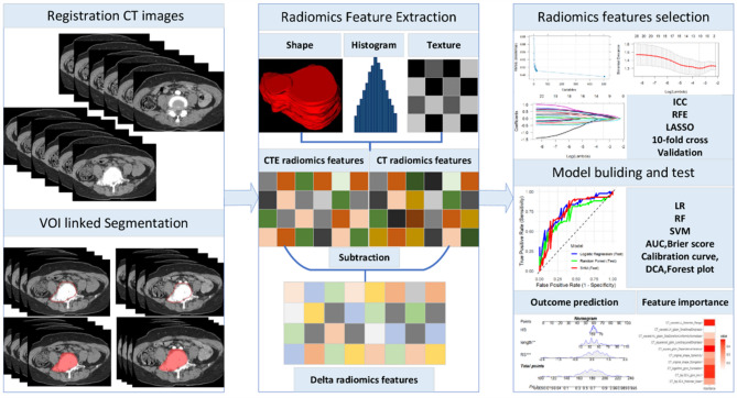
Methods: Patients diagnosed with spinal tumors who underwent spinal tumor resection surgery were retrospectively recruited. CT, CTE, delta, and clinical models based on CT native phase, CT arterial phase images, and clinical factors were constructed using 10-fold cross-validation and logistic regression (LR), random forest (RF), and support vector machine (SVM) in the training cohort. Receiver operating characteristic (ROC) curves, integrated discrimination improvement (IDI), accuracy, sensitivity, specificity, positive predictive value, and negative predictive value were used to evaluate and compare the diagnostic performance of these models.
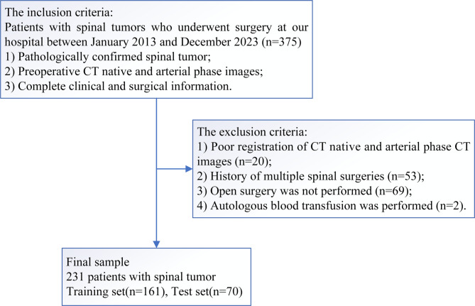
Results: 231 patients were randomly divided into training (n = 161) and test (n = 70) cohorts, comprising 146 IBT and 85 no-IBT patients, 35 IMB and 196 no-IMB patients, respectively. The delta model performed best in predicting IBT and IMB risk, with better predictive ability than the clinical model (IDI = 0.11-0.13 for IBT, and IDI = 0.02-0.08 for IMB, p < 0.05, respectively). Calibration curves indicated that the predicted probabilities of IBT and IMB in the model did not differ significantly from the actual probabilities (p > 0.05).
Conclusion: The CT delta model we constructed may be a valuable tool to improve risk stratification before spinal tumor surgery, thus contributing to preoperative planning and improving patient prognosis.
Cancer ImagingONCOLOGY-RADIOLOGY, NUCLEAR MEDICINE & MEDICAL IMAGING
CiteScore
7.00
自引率
0.00%
发文量
66
审稿时长
>12 weeks
期刊介绍:
Cancer Imaging is an open access, peer-reviewed journal publishing original articles, reviews and editorials written by expert international radiologists working in oncology.
The journal encompasses CT, MR, PET, ultrasound, radionuclide and multimodal imaging in all kinds of malignant tumours, plus new developments, techniques and innovations. Topics of interest include:
Breast Imaging
Chest
Complications of treatment
Ear, Nose & Throat
Gastrointestinal
Hepatobiliary & Pancreatic
Imaging biomarkers
Interventional
Lymphoma
Measurement of tumour response
Molecular functional imaging
Musculoskeletal
Neuro oncology
Nuclear Medicine
Paediatric.

 求助内容:
求助内容: 应助结果提醒方式:


