{"title":"尼古丁改变星形胶质细胞的细胞活性和mRNA表达模式。","authors":"Leslie Sewell, James J Cray","doi":"10.1371/journal.pone.0325529","DOIUrl":null,"url":null,"abstract":"<p><p>Nicotine exposure during neural development presents a significant public health concern. Nicotine, the primary addictive component of tobacco, influences the central nervous system by interacting with various cell types, including the glial cell termed astrocytes. Astrocytes are cells that are critical for supporting neurons, regulating neurotransmitter balance, and managing neuroinflammation. This current study explored nicotine's effects on astrocytes, examining cellular activity and gene expression within an acute exposure period. Murine C8D1A astrocytic (garnered as a cell line from postnatal day 8 tissue) cells were treated with nicotine (0-500 ng/mL) in vitro, with assays measuring cell viability and apoptosis at 12, 18, 24, and 48 hours to establish a critical concentration gradient for nicotine. Nicotine exposure increased astrocyte viability at later time points (24 and 48 hours), while apoptosis rose initially but declined over time allowing for the establishment of pharmacologically and clinically relevant nicotine concentrations of 25,50 and 100ng/ml for subsequent experiments. Real-time quantitative PCR revealed that nicotine influenced inflammatory signaling, with pro-inflammatory (A1) markers (IL-6, IFNγ, TNFα) increasing in a dose- and time-dependent manner, while anti-inflammatory (A2) markers (ARG1, IL-10, TGFβ) displayed a more complex pattern after nicotine exposures to astrocytes. These results suggest that nicotine disrupts astrocyte function and inflammatory balance, which may contribute to neurodevelopmental disruptions and heightened neuroinflammatory risks in adults. Further research is needed to investigate the prolonged impact of nicotine on brain health, addiction, and associated neurological conditions.</p>","PeriodicalId":20189,"journal":{"name":"PLoS ONE","volume":"20 6","pages":"e0325529"},"PeriodicalIF":2.6000,"publicationDate":"2025-06-20","publicationTypes":"Journal Article","fieldsOfStudy":null,"isOpenAccess":false,"openAccessPdf":"https://www.ncbi.nlm.nih.gov/pmc/articles/PMC12180639/pdf/","citationCount":"0","resultStr":"{\"title\":\"Nicotine alters cellular activity and mRNA expression of patterns of Astrocytes.\",\"authors\":\"Leslie Sewell, James J Cray\",\"doi\":\"10.1371/journal.pone.0325529\",\"DOIUrl\":null,\"url\":null,\"abstract\":\"<p><p>Nicotine exposure during neural development presents a significant public health concern. Nicotine, the primary addictive component of tobacco, influences the central nervous system by interacting with various cell types, including the glial cell termed astrocytes. Astrocytes are cells that are critical for supporting neurons, regulating neurotransmitter balance, and managing neuroinflammation. This current study explored nicotine's effects on astrocytes, examining cellular activity and gene expression within an acute exposure period. Murine C8D1A astrocytic (garnered as a cell line from postnatal day 8 tissue) cells were treated with nicotine (0-500 ng/mL) in vitro, with assays measuring cell viability and apoptosis at 12, 18, 24, and 48 hours to establish a critical concentration gradient for nicotine. Nicotine exposure increased astrocyte viability at later time points (24 and 48 hours), while apoptosis rose initially but declined over time allowing for the establishment of pharmacologically and clinically relevant nicotine concentrations of 25,50 and 100ng/ml for subsequent experiments. Real-time quantitative PCR revealed that nicotine influenced inflammatory signaling, with pro-inflammatory (A1) markers (IL-6, IFNγ, TNFα) increasing in a dose- and time-dependent manner, while anti-inflammatory (A2) markers (ARG1, IL-10, TGFβ) displayed a more complex pattern after nicotine exposures to astrocytes. These results suggest that nicotine disrupts astrocyte function and inflammatory balance, which may contribute to neurodevelopmental disruptions and heightened neuroinflammatory risks in adults. Further research is needed to investigate the prolonged impact of nicotine on brain health, addiction, and associated neurological conditions.</p>\",\"PeriodicalId\":20189,\"journal\":{\"name\":\"PLoS ONE\",\"volume\":\"20 6\",\"pages\":\"e0325529\"},\"PeriodicalIF\":2.6000,\"publicationDate\":\"2025-06-20\",\"publicationTypes\":\"Journal Article\",\"fieldsOfStudy\":null,\"isOpenAccess\":false,\"openAccessPdf\":\"https://www.ncbi.nlm.nih.gov/pmc/articles/PMC12180639/pdf/\",\"citationCount\":\"0\",\"resultStr\":null,\"platform\":\"Semanticscholar\",\"paperid\":null,\"PeriodicalName\":\"PLoS ONE\",\"FirstCategoryId\":\"103\",\"ListUrlMain\":\"https://doi.org/10.1371/journal.pone.0325529\",\"RegionNum\":3,\"RegionCategory\":\"综合性期刊\",\"ArticlePicture\":[],\"TitleCN\":null,\"AbstractTextCN\":null,\"PMCID\":null,\"EPubDate\":\"2025/1/1 0:00:00\",\"PubModel\":\"eCollection\",\"JCR\":\"Q1\",\"JCRName\":\"MULTIDISCIPLINARY SCIENCES\",\"Score\":null,\"Total\":0}","platform":"Semanticscholar","paperid":null,"PeriodicalName":"PLoS ONE","FirstCategoryId":"103","ListUrlMain":"https://doi.org/10.1371/journal.pone.0325529","RegionNum":3,"RegionCategory":"综合性期刊","ArticlePicture":[],"TitleCN":null,"AbstractTextCN":null,"PMCID":null,"EPubDate":"2025/1/1 0:00:00","PubModel":"eCollection","JCR":"Q1","JCRName":"MULTIDISCIPLINARY SCIENCES","Score":null,"Total":0}
Nicotine alters cellular activity and mRNA expression of patterns of Astrocytes.
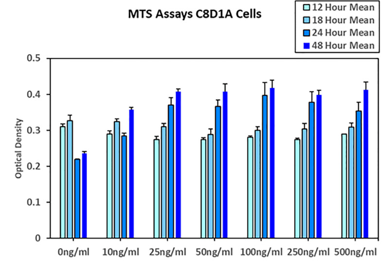
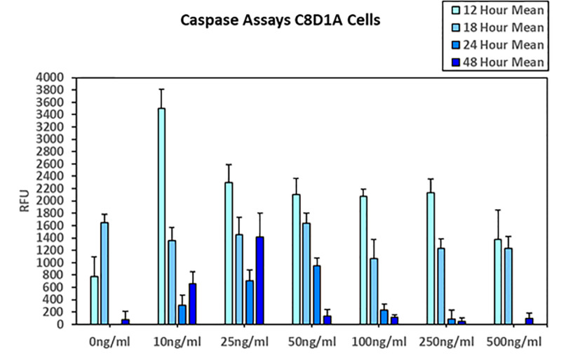
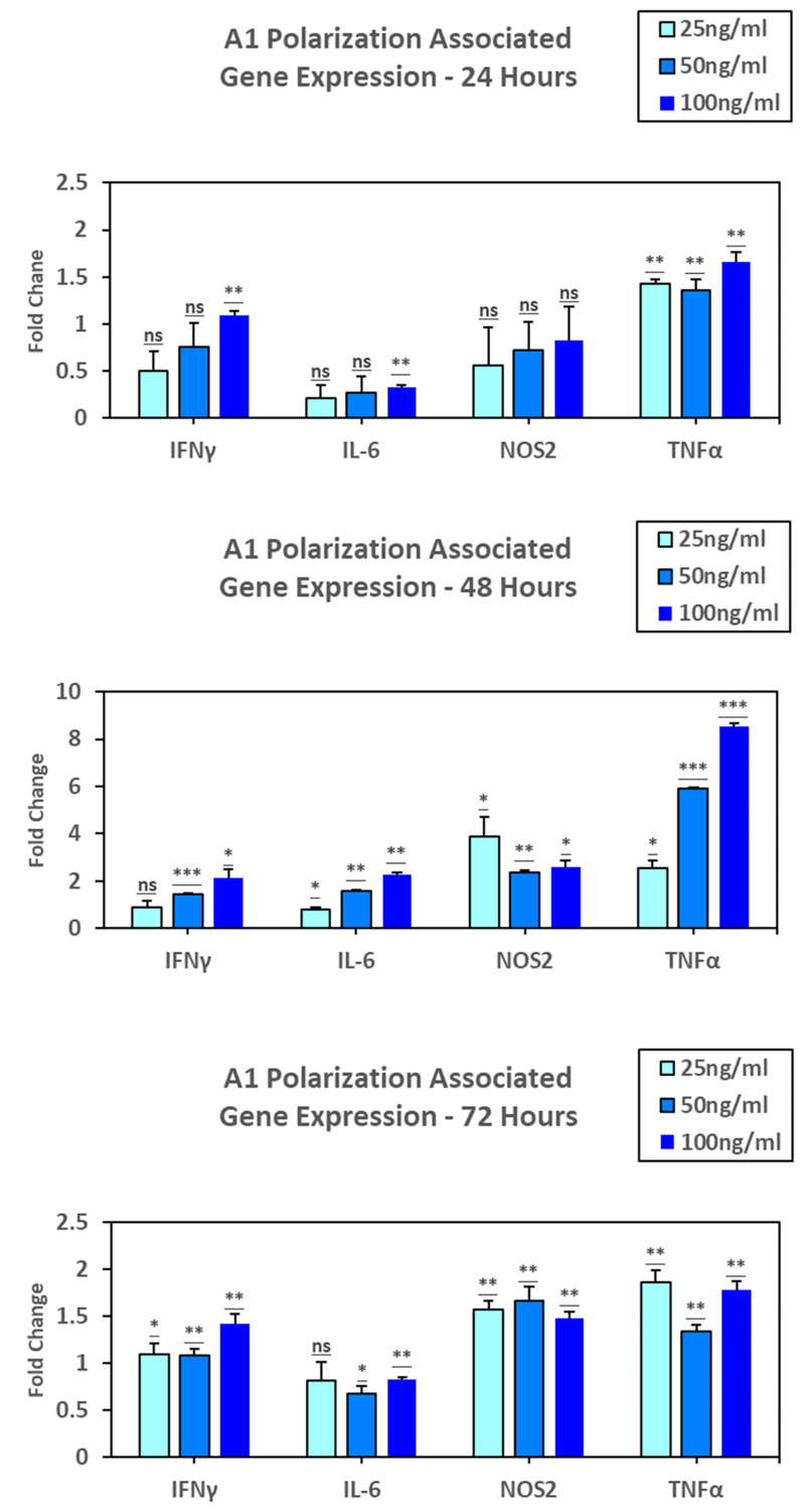
Nicotine exposure during neural development presents a significant public health concern. Nicotine, the primary addictive component of tobacco, influences the central nervous system by interacting with various cell types, including the glial cell termed astrocytes. Astrocytes are cells that are critical for supporting neurons, regulating neurotransmitter balance, and managing neuroinflammation. This current study explored nicotine's effects on astrocytes, examining cellular activity and gene expression within an acute exposure period. Murine C8D1A astrocytic (garnered as a cell line from postnatal day 8 tissue) cells were treated with nicotine (0-500 ng/mL) in vitro, with assays measuring cell viability and apoptosis at 12, 18, 24, and 48 hours to establish a critical concentration gradient for nicotine. Nicotine exposure increased astrocyte viability at later time points (24 and 48 hours), while apoptosis rose initially but declined over time allowing for the establishment of pharmacologically and clinically relevant nicotine concentrations of 25,50 and 100ng/ml for subsequent experiments. Real-time quantitative PCR revealed that nicotine influenced inflammatory signaling, with pro-inflammatory (A1) markers (IL-6, IFNγ, TNFα) increasing in a dose- and time-dependent manner, while anti-inflammatory (A2) markers (ARG1, IL-10, TGFβ) displayed a more complex pattern after nicotine exposures to astrocytes. These results suggest that nicotine disrupts astrocyte function and inflammatory balance, which may contribute to neurodevelopmental disruptions and heightened neuroinflammatory risks in adults. Further research is needed to investigate the prolonged impact of nicotine on brain health, addiction, and associated neurological conditions.
期刊介绍:
PLOS ONE is an international, peer-reviewed, open-access, online publication. PLOS ONE welcomes reports on primary research from any scientific discipline. It provides:
* Open-access—freely accessible online, authors retain copyright
* Fast publication times
* Peer review by expert, practicing researchers
* Post-publication tools to indicate quality and impact
* Community-based dialogue on articles
* Worldwide media coverage

 求助内容:
求助内容: 应助结果提醒方式:


