Mauricio Soto-Subiabre, Victor Mayoral, Lidia Valencia-Muntalà, Carlos González, Carmen Gómez-Vaquero
{"title":"椎体脆性骨折患者放射学高度恶化及其相关因素。","authors":"Mauricio Soto-Subiabre, Victor Mayoral, Lidia Valencia-Muntalà, Carlos González, Carmen Gómez-Vaquero","doi":"10.11005/jbm.25.835","DOIUrl":null,"url":null,"abstract":"<p><strong>Background: </strong>To investigate the contribution of radiological characteristics of baseline fragility vertebral fractures (FVF) and clinical characteristics to the development of radiological worsening (RW).</p><p><strong>Methods: </strong>Patients were recruited between 2015 and 2018. The primary outcome was the identification of RW in a radiological second image, defined as the progression of prevalent FVF, new FVF, or both. Data on fracture risk fractures, bone mineral density, analgesia requirements, and antiosteoporosis treatment were recorded. The radiological features of baseline FVF included fracture number, location(s), severity grade (Genant method), kyphosis angle, and spine index deformity.</p><p><strong>Results: </strong>A total of 223 patients with at least one follow-up radiological evaluation were included. Another 199 patients had no radiological follow-up. Of those with follow-up, 69% presented RW, accounting for 36.5% of the total cohort (422 patients). The incidence rate of RW was 73.8/1,000 patient-years. Among those with RW, 61% showed progression of FVF, 27% developed new FVF, and 12% had both. The multivariate analysis demonstrated that multiple FVF and worse grades of FVF at baseline were variables significantly associated with RW. Baseline characteristics of FVF that increased the risk of RW by progression of FVF was grade 1 (odds ratio [OR], 3.22; 95% confidence interval [CI], 1.47-7.02) and grade 2 (OR, 1.97; 95% CI, 1.05-3.68) and by new FVF was grade 3 (OR, 3.19; 95% CI, 1.39-7.33) FVF.</p><p><strong>Conclusions: </strong>Approximately one-third of patients with FVF experienced RW, with progression of FVF being the most common event. A higher number of FVF and a greater severity at baseline are associated with RW.</p>","PeriodicalId":15070,"journal":{"name":"Journal of Bone Metabolism","volume":"32 2","pages":"143-154"},"PeriodicalIF":0.0000,"publicationDate":"2025-05-01","publicationTypes":"Journal Article","fieldsOfStudy":null,"isOpenAccess":false,"openAccessPdf":"https://www.ncbi.nlm.nih.gov/pmc/articles/PMC12183356/pdf/","citationCount":"0","resultStr":"{\"title\":\"High Radiological Worsening in Patients with Vertebral Fragility Fractures and the Associated Factors.\",\"authors\":\"Mauricio Soto-Subiabre, Victor Mayoral, Lidia Valencia-Muntalà, Carlos González, Carmen Gómez-Vaquero\",\"doi\":\"10.11005/jbm.25.835\",\"DOIUrl\":null,\"url\":null,\"abstract\":\"<p><strong>Background: </strong>To investigate the contribution of radiological characteristics of baseline fragility vertebral fractures (FVF) and clinical characteristics to the development of radiological worsening (RW).</p><p><strong>Methods: </strong>Patients were recruited between 2015 and 2018. The primary outcome was the identification of RW in a radiological second image, defined as the progression of prevalent FVF, new FVF, or both. Data on fracture risk fractures, bone mineral density, analgesia requirements, and antiosteoporosis treatment were recorded. The radiological features of baseline FVF included fracture number, location(s), severity grade (Genant method), kyphosis angle, and spine index deformity.</p><p><strong>Results: </strong>A total of 223 patients with at least one follow-up radiological evaluation were included. Another 199 patients had no radiological follow-up. Of those with follow-up, 69% presented RW, accounting for 36.5% of the total cohort (422 patients). The incidence rate of RW was 73.8/1,000 patient-years. Among those with RW, 61% showed progression of FVF, 27% developed new FVF, and 12% had both. The multivariate analysis demonstrated that multiple FVF and worse grades of FVF at baseline were variables significantly associated with RW. Baseline characteristics of FVF that increased the risk of RW by progression of FVF was grade 1 (odds ratio [OR], 3.22; 95% confidence interval [CI], 1.47-7.02) and grade 2 (OR, 1.97; 95% CI, 1.05-3.68) and by new FVF was grade 3 (OR, 3.19; 95% CI, 1.39-7.33) FVF.</p><p><strong>Conclusions: </strong>Approximately one-third of patients with FVF experienced RW, with progression of FVF being the most common event. A higher number of FVF and a greater severity at baseline are associated with RW.</p>\",\"PeriodicalId\":15070,\"journal\":{\"name\":\"Journal of Bone Metabolism\",\"volume\":\"32 2\",\"pages\":\"143-154\"},\"PeriodicalIF\":0.0000,\"publicationDate\":\"2025-05-01\",\"publicationTypes\":\"Journal Article\",\"fieldsOfStudy\":null,\"isOpenAccess\":false,\"openAccessPdf\":\"https://www.ncbi.nlm.nih.gov/pmc/articles/PMC12183356/pdf/\",\"citationCount\":\"0\",\"resultStr\":null,\"platform\":\"Semanticscholar\",\"paperid\":null,\"PeriodicalName\":\"Journal of Bone Metabolism\",\"FirstCategoryId\":\"1085\",\"ListUrlMain\":\"https://doi.org/10.11005/jbm.25.835\",\"RegionNum\":0,\"RegionCategory\":null,\"ArticlePicture\":[],\"TitleCN\":null,\"AbstractTextCN\":null,\"PMCID\":null,\"EPubDate\":\"2025/5/31 0:00:00\",\"PubModel\":\"Epub\",\"JCR\":\"Q2\",\"JCRName\":\"Medicine\",\"Score\":null,\"Total\":0}","platform":"Semanticscholar","paperid":null,"PeriodicalName":"Journal of Bone Metabolism","FirstCategoryId":"1085","ListUrlMain":"https://doi.org/10.11005/jbm.25.835","RegionNum":0,"RegionCategory":null,"ArticlePicture":[],"TitleCN":null,"AbstractTextCN":null,"PMCID":null,"EPubDate":"2025/5/31 0:00:00","PubModel":"Epub","JCR":"Q2","JCRName":"Medicine","Score":null,"Total":0}
High Radiological Worsening in Patients with Vertebral Fragility Fractures and the Associated Factors.
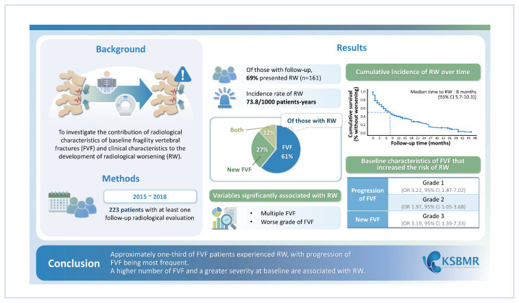
Background: To investigate the contribution of radiological characteristics of baseline fragility vertebral fractures (FVF) and clinical characteristics to the development of radiological worsening (RW).
Methods: Patients were recruited between 2015 and 2018. The primary outcome was the identification of RW in a radiological second image, defined as the progression of prevalent FVF, new FVF, or both. Data on fracture risk fractures, bone mineral density, analgesia requirements, and antiosteoporosis treatment were recorded. The radiological features of baseline FVF included fracture number, location(s), severity grade (Genant method), kyphosis angle, and spine index deformity.
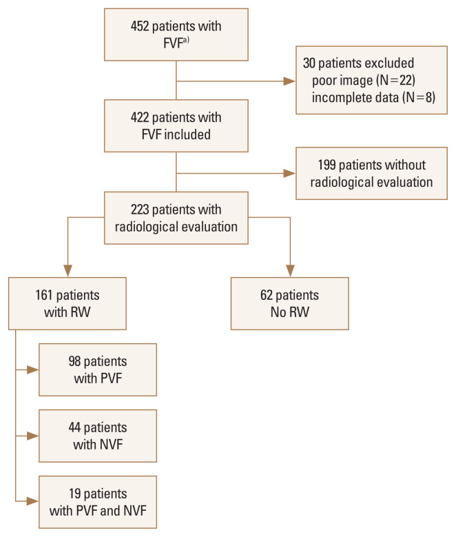
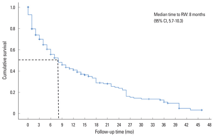
Results: A total of 223 patients with at least one follow-up radiological evaluation were included. Another 199 patients had no radiological follow-up. Of those with follow-up, 69% presented RW, accounting for 36.5% of the total cohort (422 patients). The incidence rate of RW was 73.8/1,000 patient-years. Among those with RW, 61% showed progression of FVF, 27% developed new FVF, and 12% had both. The multivariate analysis demonstrated that multiple FVF and worse grades of FVF at baseline were variables significantly associated with RW. Baseline characteristics of FVF that increased the risk of RW by progression of FVF was grade 1 (odds ratio [OR], 3.22; 95% confidence interval [CI], 1.47-7.02) and grade 2 (OR, 1.97; 95% CI, 1.05-3.68) and by new FVF was grade 3 (OR, 3.19; 95% CI, 1.39-7.33) FVF.
Conclusions: Approximately one-third of patients with FVF experienced RW, with progression of FVF being the most common event. A higher number of FVF and a greater severity at baseline are associated with RW.

 求助内容:
求助内容: 应助结果提醒方式:


