{"title":"超声直方图分析评估COVID-19患者睾丸实质的病例对照研究","authors":"Mehmet Demir, Hatem Kazımoğlu","doi":"10.1590/1806-9282.20250074","DOIUrl":null,"url":null,"abstract":"<p><strong>Objective: </strong>Uncontrolled activation of immune cells, such as lymphocytes and macrophages, can result in heightened inflammatory cytokine production, leading to oxidative stress and cell death in various organs, including the brain and testes. The systemic effects of COVID-19 are not limited to the respiratory system and can also affect other organs. The aim of this study was to focus on the possible effects of COVID-19 infection on the testicles in male patients.</p><p><strong>Methods: </strong>Ultrasound images of 50 patients who were shown to have COVID-19 infection by polymerase chain reaction method and 50 healthy volunteers who did not have any complaints and were polymerase chain reaction negative were evaluated retrospectively. While forming the patient and control groups, people who smoked or had diseases such as varicocele, which caused testicular parenchyma to be affected, were excluded from the study. Tissue histogram analysis was performed on the images obtained, including the entire testicular parenchyma, and radiomics were obtained. The Mann-Whitney U test and independent samples t-test were used to compare parametric and non-parametric results in independent groups.</p><p><strong>Results: </strong>The average age of the patient group and control group was 32.13±7.3 years and 31.23±7.9 years, respectively, and no statistically significant difference was detected (p>0.05). When testicular volumes in the patient and control groups were compared, no statistically significant difference was detected (p=0.58). Statistically significant differences were found between the two groups in the radiomics examined using skewness of histogram, zone-size non-uniformity of gray-level size-zone matrix, information measure of correlation of gray-level co-occurrence matrix, run percentage of gray-level run-length matrix, high gray-level run emphasis of gray-level run-length matrix, and kurtosis of histogram (p<0.05). There was no significant difference in other parameters.</p><p><strong>Conclusion: </strong>Ultrasound histogram analysis may be useful in showing heterogeneity, which is an indicator of the impact on the testicular parenchyma in people with COVID-19 infection. Thus, in addition to other clinical findings and laboratory parameters, ultrasound histogram analysis may be helpful in demonstrating testicular parenchymal involvement in the follow-up of patients after COVID-19.</p>","PeriodicalId":94194,"journal":{"name":"Revista da Associacao Medica Brasileira (1992)","volume":"71 5","pages":"e20250074"},"PeriodicalIF":1.3000,"publicationDate":"2025-06-16","publicationTypes":"Journal Article","fieldsOfStudy":null,"isOpenAccess":false,"openAccessPdf":"https://www.ncbi.nlm.nih.gov/pmc/articles/PMC12172521/pdf/","citationCount":"0","resultStr":"{\"title\":\"Assessment of testicular parenchyma in COVID-19 patients: a case-control study using ultrasound histogram analysis.\",\"authors\":\"Mehmet Demir, Hatem Kazımoğlu\",\"doi\":\"10.1590/1806-9282.20250074\",\"DOIUrl\":null,\"url\":null,\"abstract\":\"<p><strong>Objective: </strong>Uncontrolled activation of immune cells, such as lymphocytes and macrophages, can result in heightened inflammatory cytokine production, leading to oxidative stress and cell death in various organs, including the brain and testes. The systemic effects of COVID-19 are not limited to the respiratory system and can also affect other organs. The aim of this study was to focus on the possible effects of COVID-19 infection on the testicles in male patients.</p><p><strong>Methods: </strong>Ultrasound images of 50 patients who were shown to have COVID-19 infection by polymerase chain reaction method and 50 healthy volunteers who did not have any complaints and were polymerase chain reaction negative were evaluated retrospectively. While forming the patient and control groups, people who smoked or had diseases such as varicocele, which caused testicular parenchyma to be affected, were excluded from the study. Tissue histogram analysis was performed on the images obtained, including the entire testicular parenchyma, and radiomics were obtained. The Mann-Whitney U test and independent samples t-test were used to compare parametric and non-parametric results in independent groups.</p><p><strong>Results: </strong>The average age of the patient group and control group was 32.13±7.3 years and 31.23±7.9 years, respectively, and no statistically significant difference was detected (p>0.05). When testicular volumes in the patient and control groups were compared, no statistically significant difference was detected (p=0.58). Statistically significant differences were found between the two groups in the radiomics examined using skewness of histogram, zone-size non-uniformity of gray-level size-zone matrix, information measure of correlation of gray-level co-occurrence matrix, run percentage of gray-level run-length matrix, high gray-level run emphasis of gray-level run-length matrix, and kurtosis of histogram (p<0.05). There was no significant difference in other parameters.</p><p><strong>Conclusion: </strong>Ultrasound histogram analysis may be useful in showing heterogeneity, which is an indicator of the impact on the testicular parenchyma in people with COVID-19 infection. Thus, in addition to other clinical findings and laboratory parameters, ultrasound histogram analysis may be helpful in demonstrating testicular parenchymal involvement in the follow-up of patients after COVID-19.</p>\",\"PeriodicalId\":94194,\"journal\":{\"name\":\"Revista da Associacao Medica Brasileira (1992)\",\"volume\":\"71 5\",\"pages\":\"e20250074\"},\"PeriodicalIF\":1.3000,\"publicationDate\":\"2025-06-16\",\"publicationTypes\":\"Journal Article\",\"fieldsOfStudy\":null,\"isOpenAccess\":false,\"openAccessPdf\":\"https://www.ncbi.nlm.nih.gov/pmc/articles/PMC12172521/pdf/\",\"citationCount\":\"0\",\"resultStr\":null,\"platform\":\"Semanticscholar\",\"paperid\":null,\"PeriodicalName\":\"Revista da Associacao Medica Brasileira (1992)\",\"FirstCategoryId\":\"1085\",\"ListUrlMain\":\"https://doi.org/10.1590/1806-9282.20250074\",\"RegionNum\":0,\"RegionCategory\":null,\"ArticlePicture\":[],\"TitleCN\":null,\"AbstractTextCN\":null,\"PMCID\":null,\"EPubDate\":\"2025/1/1 0:00:00\",\"PubModel\":\"eCollection\",\"JCR\":\"\",\"JCRName\":\"\",\"Score\":null,\"Total\":0}","platform":"Semanticscholar","paperid":null,"PeriodicalName":"Revista da Associacao Medica Brasileira (1992)","FirstCategoryId":"1085","ListUrlMain":"https://doi.org/10.1590/1806-9282.20250074","RegionNum":0,"RegionCategory":null,"ArticlePicture":[],"TitleCN":null,"AbstractTextCN":null,"PMCID":null,"EPubDate":"2025/1/1 0:00:00","PubModel":"eCollection","JCR":"","JCRName":"","Score":null,"Total":0}
Assessment of testicular parenchyma in COVID-19 patients: a case-control study using ultrasound histogram analysis.
Objective: Uncontrolled activation of immune cells, such as lymphocytes and macrophages, can result in heightened inflammatory cytokine production, leading to oxidative stress and cell death in various organs, including the brain and testes. The systemic effects of COVID-19 are not limited to the respiratory system and can also affect other organs. The aim of this study was to focus on the possible effects of COVID-19 infection on the testicles in male patients.
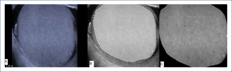
Methods: Ultrasound images of 50 patients who were shown to have COVID-19 infection by polymerase chain reaction method and 50 healthy volunteers who did not have any complaints and were polymerase chain reaction negative were evaluated retrospectively. While forming the patient and control groups, people who smoked or had diseases such as varicocele, which caused testicular parenchyma to be affected, were excluded from the study. Tissue histogram analysis was performed on the images obtained, including the entire testicular parenchyma, and radiomics were obtained. The Mann-Whitney U test and independent samples t-test were used to compare parametric and non-parametric results in independent groups.
Results: The average age of the patient group and control group was 32.13±7.3 years and 31.23±7.9 years, respectively, and no statistically significant difference was detected (p>0.05). When testicular volumes in the patient and control groups were compared, no statistically significant difference was detected (p=0.58). Statistically significant differences were found between the two groups in the radiomics examined using skewness of histogram, zone-size non-uniformity of gray-level size-zone matrix, information measure of correlation of gray-level co-occurrence matrix, run percentage of gray-level run-length matrix, high gray-level run emphasis of gray-level run-length matrix, and kurtosis of histogram (p<0.05). There was no significant difference in other parameters.
Conclusion: Ultrasound histogram analysis may be useful in showing heterogeneity, which is an indicator of the impact on the testicular parenchyma in people with COVID-19 infection. Thus, in addition to other clinical findings and laboratory parameters, ultrasound histogram analysis may be helpful in demonstrating testicular parenchymal involvement in the follow-up of patients after COVID-19.

 求助内容:
求助内容: 应助结果提醒方式:


