{"title":"基于2.5D磁共振成像的腮腺肿瘤深度学习鉴别诊断","authors":"Wenfeng Mai, Xiaole Fan, Lingtao Zhang, Jian Li, Liting Chen, Xiaoyu Hua, Dong Zhang, Hengguo Li, Minxiang Cai, Changzheng Shi, Xiangning Liu","doi":"10.1080/07853890.2025.2520401","DOIUrl":null,"url":null,"abstract":"<p><strong>Purpose: </strong>Accurate preoperative diagnosis of parotid gland tumors (PGTs) is crucial for surgical planning since malignant tumors require more extensive excision. Though fine-needle aspiration biopsy is the diagnostic gold standard, its sensitivity in detecting malignancies is limited. While Deep learning (DL) models based on magnetic resonance imaging (MRI) are common in medicine, they are less studied for parotid gland tumors. This study used a 2.5D imaging approach (Incorporating Inter-Slice Information) to train a DL model to differentiate between benign and malignant PGTs.</p><p><strong>Methods: </strong>This retrospective study included 122 parotid tumor patients, using MRI and clinical features to build predictive models. In the traditional model, univariate analysis identified statistically significant features, which were then used in multivariate logistic regression to determine independent predictors. The model was built using four-fold cross-validation. The deep learning model was trained using 2D and 2.5D imaging approaches, with a transformer-based architecture employed for transfer learning. The model's performance was evaluated using the area under the receiver operating characteristic curve (AUC) and confusion matrix metrics.</p><p><strong>Results: </strong>In the traditional model, boundary and peritumoral invasion were identified as independent predictors for PGTs, and the model was constructed based on these features. The model achieved an AUC of 0.79 but demonstrated low sensitivity (0.54). In contrast, the DL model based on 2.5D T2 fat-suppressed images showed superior performance, with an AUC of 0.86 and a sensitivity of 0.78.</p><p><strong>Conclusion: </strong>The 2.5D imaging technique, when integrated with a transformer-based transfer learning model, demonstrates significant efficacy in differentiating between PGTs.</p>","PeriodicalId":93874,"journal":{"name":"Annals of medicine","volume":"57 1","pages":"2520401"},"PeriodicalIF":4.3000,"publicationDate":"2025-12-01","publicationTypes":"Journal Article","fieldsOfStudy":null,"isOpenAccess":false,"openAccessPdf":"https://www.ncbi.nlm.nih.gov/pmc/articles/PMC12180351/pdf/","citationCount":"0","resultStr":"{\"title\":\"Deep learning for differential diagnosis of parotid tumors based on 2.5D magnetic resonance imaging.\",\"authors\":\"Wenfeng Mai, Xiaole Fan, Lingtao Zhang, Jian Li, Liting Chen, Xiaoyu Hua, Dong Zhang, Hengguo Li, Minxiang Cai, Changzheng Shi, Xiangning Liu\",\"doi\":\"10.1080/07853890.2025.2520401\",\"DOIUrl\":null,\"url\":null,\"abstract\":\"<p><strong>Purpose: </strong>Accurate preoperative diagnosis of parotid gland tumors (PGTs) is crucial for surgical planning since malignant tumors require more extensive excision. Though fine-needle aspiration biopsy is the diagnostic gold standard, its sensitivity in detecting malignancies is limited. While Deep learning (DL) models based on magnetic resonance imaging (MRI) are common in medicine, they are less studied for parotid gland tumors. This study used a 2.5D imaging approach (Incorporating Inter-Slice Information) to train a DL model to differentiate between benign and malignant PGTs.</p><p><strong>Methods: </strong>This retrospective study included 122 parotid tumor patients, using MRI and clinical features to build predictive models. In the traditional model, univariate analysis identified statistically significant features, which were then used in multivariate logistic regression to determine independent predictors. The model was built using four-fold cross-validation. The deep learning model was trained using 2D and 2.5D imaging approaches, with a transformer-based architecture employed for transfer learning. The model's performance was evaluated using the area under the receiver operating characteristic curve (AUC) and confusion matrix metrics.</p><p><strong>Results: </strong>In the traditional model, boundary and peritumoral invasion were identified as independent predictors for PGTs, and the model was constructed based on these features. The model achieved an AUC of 0.79 but demonstrated low sensitivity (0.54). In contrast, the DL model based on 2.5D T2 fat-suppressed images showed superior performance, with an AUC of 0.86 and a sensitivity of 0.78.</p><p><strong>Conclusion: </strong>The 2.5D imaging technique, when integrated with a transformer-based transfer learning model, demonstrates significant efficacy in differentiating between PGTs.</p>\",\"PeriodicalId\":93874,\"journal\":{\"name\":\"Annals of medicine\",\"volume\":\"57 1\",\"pages\":\"2520401\"},\"PeriodicalIF\":4.3000,\"publicationDate\":\"2025-12-01\",\"publicationTypes\":\"Journal Article\",\"fieldsOfStudy\":null,\"isOpenAccess\":false,\"openAccessPdf\":\"https://www.ncbi.nlm.nih.gov/pmc/articles/PMC12180351/pdf/\",\"citationCount\":\"0\",\"resultStr\":null,\"platform\":\"Semanticscholar\",\"paperid\":null,\"PeriodicalName\":\"Annals of medicine\",\"FirstCategoryId\":\"1085\",\"ListUrlMain\":\"https://doi.org/10.1080/07853890.2025.2520401\",\"RegionNum\":0,\"RegionCategory\":null,\"ArticlePicture\":[],\"TitleCN\":null,\"AbstractTextCN\":null,\"PMCID\":null,\"EPubDate\":\"2025/6/18 0:00:00\",\"PubModel\":\"Epub\",\"JCR\":\"\",\"JCRName\":\"\",\"Score\":null,\"Total\":0}","platform":"Semanticscholar","paperid":null,"PeriodicalName":"Annals of medicine","FirstCategoryId":"1085","ListUrlMain":"https://doi.org/10.1080/07853890.2025.2520401","RegionNum":0,"RegionCategory":null,"ArticlePicture":[],"TitleCN":null,"AbstractTextCN":null,"PMCID":null,"EPubDate":"2025/6/18 0:00:00","PubModel":"Epub","JCR":"","JCRName":"","Score":null,"Total":0}
Deep learning for differential diagnosis of parotid tumors based on 2.5D magnetic resonance imaging.
Purpose: Accurate preoperative diagnosis of parotid gland tumors (PGTs) is crucial for surgical planning since malignant tumors require more extensive excision. Though fine-needle aspiration biopsy is the diagnostic gold standard, its sensitivity in detecting malignancies is limited. While Deep learning (DL) models based on magnetic resonance imaging (MRI) are common in medicine, they are less studied for parotid gland tumors. This study used a 2.5D imaging approach (Incorporating Inter-Slice Information) to train a DL model to differentiate between benign and malignant PGTs.
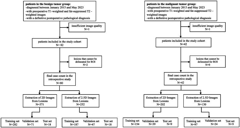
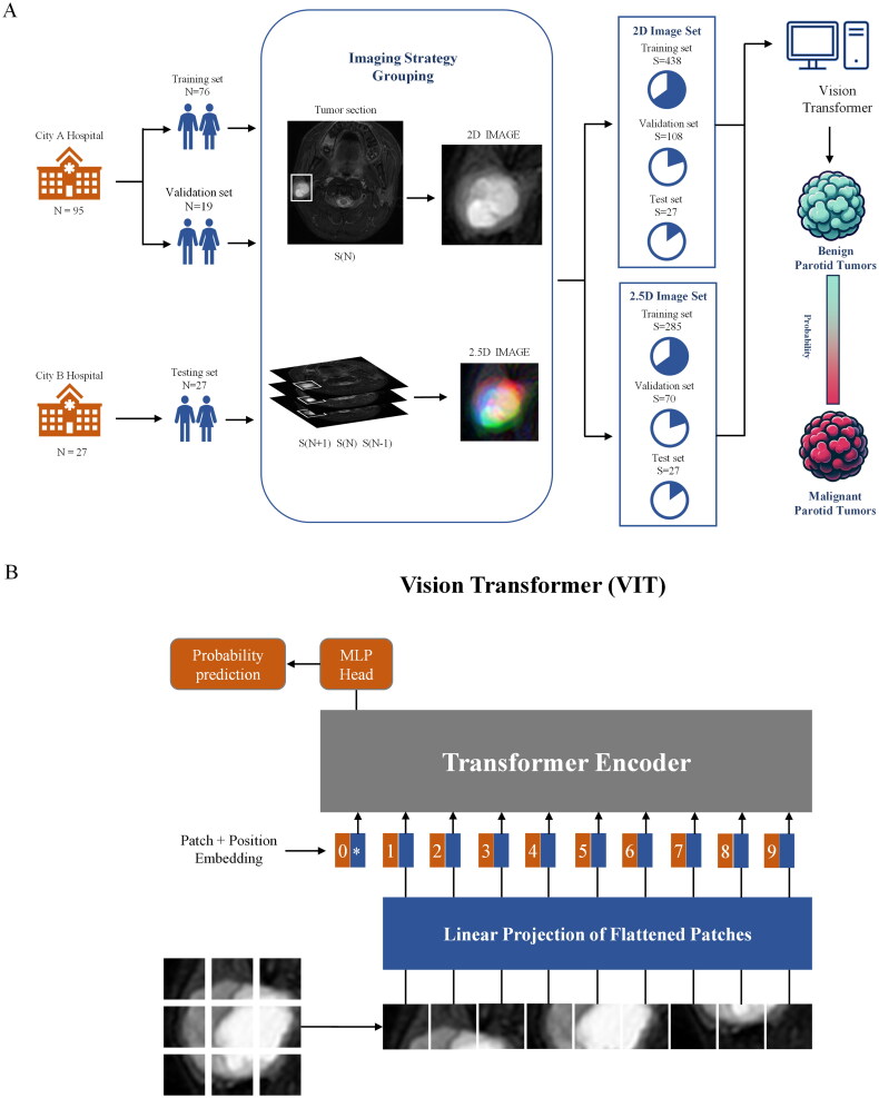
Methods: This retrospective study included 122 parotid tumor patients, using MRI and clinical features to build predictive models. In the traditional model, univariate analysis identified statistically significant features, which were then used in multivariate logistic regression to determine independent predictors. The model was built using four-fold cross-validation. The deep learning model was trained using 2D and 2.5D imaging approaches, with a transformer-based architecture employed for transfer learning. The model's performance was evaluated using the area under the receiver operating characteristic curve (AUC) and confusion matrix metrics.
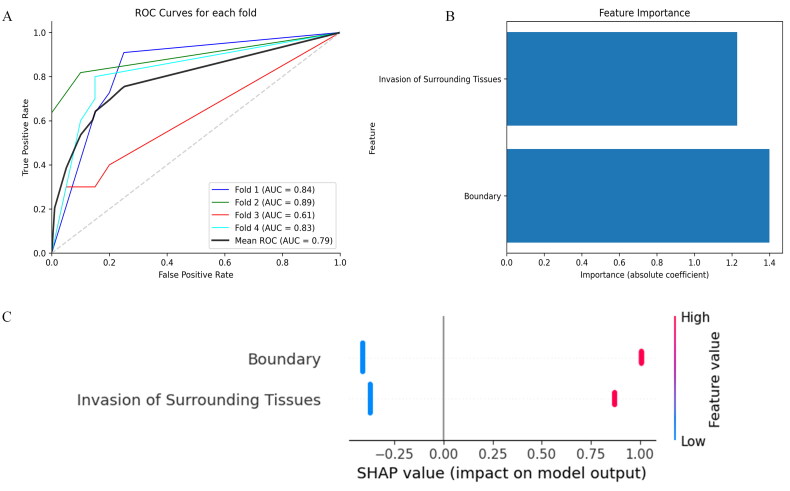
Results: In the traditional model, boundary and peritumoral invasion were identified as independent predictors for PGTs, and the model was constructed based on these features. The model achieved an AUC of 0.79 but demonstrated low sensitivity (0.54). In contrast, the DL model based on 2.5D T2 fat-suppressed images showed superior performance, with an AUC of 0.86 and a sensitivity of 0.78.
Conclusion: The 2.5D imaging technique, when integrated with a transformer-based transfer learning model, demonstrates significant efficacy in differentiating between PGTs.

 求助内容:
求助内容: 应助结果提醒方式:


