Corinna Trenker, Christian Görg, Andreas Burchert, Christina C Westhoff, Ehsan Safai Zadeh, Christoph F Dietrich, Hajo Findeisen, Christoph Mann
{"title":"髓外骨髓瘤在b超和增强超声上的表现。","authors":"Corinna Trenker, Christian Görg, Andreas Burchert, Christina C Westhoff, Ehsan Safai Zadeh, Christoph F Dietrich, Hajo Findeisen, Christoph Mann","doi":"10.15557/jou.2025.0015","DOIUrl":null,"url":null,"abstract":"<p><strong>Aim: </strong>In patients with multiple myeloma, extramedullary myeloma manifestations can occur alongside bone marrow infiltration and osseous involvement. The aim of this study was to describe extramedullary myeloma manifestations using B-mode ultrasound and contrast-enhanced ultrasound.</p><p><strong>Material and methods: </strong>Between February 2006 and 2021, a total of 21 patients with multiple myeloma and histologically or clinically proven extramedullary myeloma manifestations (<i>n</i> = 24) were included. All patients underwent B-mode ultrasound and contrast-enhanced ultrasound of extramedullary myeloma manifestations. B-mode ultrasound patterns of location, size border characteristics, and echogenicity (hypoechoic/isoechoic or hyperechoic) as well as contrast-enhanced ultrasound enhancement (hyper-, iso-, or hypoenhancement) were analyzed.</p><p><strong>Results: </strong>In most cases, extramedullary myeloma manifestations were located in the chest wall (<i>n</i> = 11; 45.8%). In all 24 cases, extramedullary myeloma manifestations were hypoechoic on B-mode ultrasound. <i>N</i> = 16 (66.6%) of extramedullary myeloma manifestations had smooth and <i>n</i> = 8 (33.3%) had irregular borders. The mean lesion size was 5.4 cm. On contrast-enhanced ultrasound, extramedullary myeloma manifestations presented arterial hyper- (<i>n</i> = 20; 83.3%) or isoenhancement (<i>n</i> = 4; 16.7%) followed by parenchymal iso- (<i>n</i> = 1; 4.2%) or hypoenhancement (<i>n</i> = 23; 95.8%). In molecular genetic analysis, every patient with reliable FISH results tested positive for at least one aberration considered \"high-risk\".</p><p><strong>Conclusion: </strong>Extramedullary myeloma manifestations were typically hypoechoic on B-mode ultrasound. On contrast-enhanced ultrasound, they presented characteristic arterial hyperenhancement followed by parenchymal washout. All patients studied for the genetic risk status were found to be \"high-risk\".</p>","PeriodicalId":45612,"journal":{"name":"Journal of Ultrasonography","volume":"25 101","pages":"20250015"},"PeriodicalIF":1.5000,"publicationDate":"2025-05-08","publicationTypes":"Journal Article","fieldsOfStudy":null,"isOpenAccess":false,"openAccessPdf":"https://www.ncbi.nlm.nih.gov/pmc/articles/PMC12175145/pdf/","citationCount":"0","resultStr":"{\"title\":\"Presentation of extramedullary myeloma manifestations on B-mode (B-US) and contrast-enhanced ultrasound (CEUS).\",\"authors\":\"Corinna Trenker, Christian Görg, Andreas Burchert, Christina C Westhoff, Ehsan Safai Zadeh, Christoph F Dietrich, Hajo Findeisen, Christoph Mann\",\"doi\":\"10.15557/jou.2025.0015\",\"DOIUrl\":null,\"url\":null,\"abstract\":\"<p><strong>Aim: </strong>In patients with multiple myeloma, extramedullary myeloma manifestations can occur alongside bone marrow infiltration and osseous involvement. The aim of this study was to describe extramedullary myeloma manifestations using B-mode ultrasound and contrast-enhanced ultrasound.</p><p><strong>Material and methods: </strong>Between February 2006 and 2021, a total of 21 patients with multiple myeloma and histologically or clinically proven extramedullary myeloma manifestations (<i>n</i> = 24) were included. All patients underwent B-mode ultrasound and contrast-enhanced ultrasound of extramedullary myeloma manifestations. B-mode ultrasound patterns of location, size border characteristics, and echogenicity (hypoechoic/isoechoic or hyperechoic) as well as contrast-enhanced ultrasound enhancement (hyper-, iso-, or hypoenhancement) were analyzed.</p><p><strong>Results: </strong>In most cases, extramedullary myeloma manifestations were located in the chest wall (<i>n</i> = 11; 45.8%). In all 24 cases, extramedullary myeloma manifestations were hypoechoic on B-mode ultrasound. <i>N</i> = 16 (66.6%) of extramedullary myeloma manifestations had smooth and <i>n</i> = 8 (33.3%) had irregular borders. The mean lesion size was 5.4 cm. On contrast-enhanced ultrasound, extramedullary myeloma manifestations presented arterial hyper- (<i>n</i> = 20; 83.3%) or isoenhancement (<i>n</i> = 4; 16.7%) followed by parenchymal iso- (<i>n</i> = 1; 4.2%) or hypoenhancement (<i>n</i> = 23; 95.8%). In molecular genetic analysis, every patient with reliable FISH results tested positive for at least one aberration considered \\\"high-risk\\\".</p><p><strong>Conclusion: </strong>Extramedullary myeloma manifestations were typically hypoechoic on B-mode ultrasound. On contrast-enhanced ultrasound, they presented characteristic arterial hyperenhancement followed by parenchymal washout. All patients studied for the genetic risk status were found to be \\\"high-risk\\\".</p>\",\"PeriodicalId\":45612,\"journal\":{\"name\":\"Journal of Ultrasonography\",\"volume\":\"25 101\",\"pages\":\"20250015\"},\"PeriodicalIF\":1.5000,\"publicationDate\":\"2025-05-08\",\"publicationTypes\":\"Journal Article\",\"fieldsOfStudy\":null,\"isOpenAccess\":false,\"openAccessPdf\":\"https://www.ncbi.nlm.nih.gov/pmc/articles/PMC12175145/pdf/\",\"citationCount\":\"0\",\"resultStr\":null,\"platform\":\"Semanticscholar\",\"paperid\":null,\"PeriodicalName\":\"Journal of Ultrasonography\",\"FirstCategoryId\":\"1085\",\"ListUrlMain\":\"https://doi.org/10.15557/jou.2025.0015\",\"RegionNum\":0,\"RegionCategory\":null,\"ArticlePicture\":[],\"TitleCN\":null,\"AbstractTextCN\":null,\"PMCID\":null,\"EPubDate\":\"2025/4/1 0:00:00\",\"PubModel\":\"eCollection\",\"JCR\":\"Q3\",\"JCRName\":\"RADIOLOGY, NUCLEAR MEDICINE & MEDICAL IMAGING\",\"Score\":null,\"Total\":0}","platform":"Semanticscholar","paperid":null,"PeriodicalName":"Journal of Ultrasonography","FirstCategoryId":"1085","ListUrlMain":"https://doi.org/10.15557/jou.2025.0015","RegionNum":0,"RegionCategory":null,"ArticlePicture":[],"TitleCN":null,"AbstractTextCN":null,"PMCID":null,"EPubDate":"2025/4/1 0:00:00","PubModel":"eCollection","JCR":"Q3","JCRName":"RADIOLOGY, NUCLEAR MEDICINE & MEDICAL IMAGING","Score":null,"Total":0}
Presentation of extramedullary myeloma manifestations on B-mode (B-US) and contrast-enhanced ultrasound (CEUS).
Aim: In patients with multiple myeloma, extramedullary myeloma manifestations can occur alongside bone marrow infiltration and osseous involvement. The aim of this study was to describe extramedullary myeloma manifestations using B-mode ultrasound and contrast-enhanced ultrasound.
Material and methods: Between February 2006 and 2021, a total of 21 patients with multiple myeloma and histologically or clinically proven extramedullary myeloma manifestations (n = 24) were included. All patients underwent B-mode ultrasound and contrast-enhanced ultrasound of extramedullary myeloma manifestations. B-mode ultrasound patterns of location, size border characteristics, and echogenicity (hypoechoic/isoechoic or hyperechoic) as well as contrast-enhanced ultrasound enhancement (hyper-, iso-, or hypoenhancement) were analyzed.
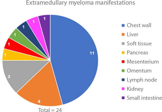
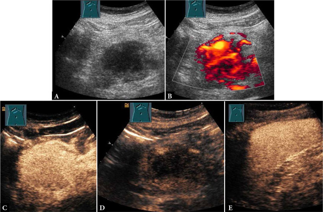
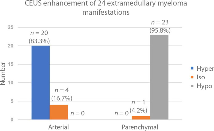
Results: In most cases, extramedullary myeloma manifestations were located in the chest wall (n = 11; 45.8%). In all 24 cases, extramedullary myeloma manifestations were hypoechoic on B-mode ultrasound. N = 16 (66.6%) of extramedullary myeloma manifestations had smooth and n = 8 (33.3%) had irregular borders. The mean lesion size was 5.4 cm. On contrast-enhanced ultrasound, extramedullary myeloma manifestations presented arterial hyper- (n = 20; 83.3%) or isoenhancement (n = 4; 16.7%) followed by parenchymal iso- (n = 1; 4.2%) or hypoenhancement (n = 23; 95.8%). In molecular genetic analysis, every patient with reliable FISH results tested positive for at least one aberration considered "high-risk".
Conclusion: Extramedullary myeloma manifestations were typically hypoechoic on B-mode ultrasound. On contrast-enhanced ultrasound, they presented characteristic arterial hyperenhancement followed by parenchymal washout. All patients studied for the genetic risk status were found to be "high-risk".

 求助内容:
求助内容: 应助结果提醒方式:


