{"title":"伊鲁替尼蛋白偶联物在大鼠胃肠道定位的免疫组织化学研究。","authors":"Hiroto Kataoka, Tetsuya Saita, Yutaro Yamamoto, Sakiko Kimura, Rintaro Sogawa, Chisato Shimanoe","doi":"10.1267/ahc.24-00053","DOIUrl":null,"url":null,"abstract":"<p><p>Ibrutinib is an oral irreversible Bruton's tyrosine kinase (BTK) inhibitor that blocks BTK activity by forming covalent bonds with the thiol group of cysteine in the ATP-binding pocket via Michael addition. However, it also reacts with a variety of off-target nonspecific proteins. In this study, we attempted to generate a specific antibody against ibrutinib and develop an immunohistochemical method to detect the ibrutinib-protein conjugates. Ibrutinib has the same amino group as the nucleobase adenine. Paraformaldehyde fixation could not fix it to the tissue via this amino group. However, ibrutinib covalently binds to proteins such as BTKs to exert its action and is therefore immobilized in tissue as ibrutinib-protein conjugates. Thus, immunohistochemistry for ibrutinib detects the location of the ibrutinib-protein conjugates, that is, the sites of covalently bound to the tissue via Michael addition. Using this immunohistochemical method, we visualized the ibrutinib-protein conjugates in the rat gastrointestinal tract (gastric body, duodenum, jejunum, ileum, and colon). This study is the first to elucidate the location of the ibrutinib-protein conjugates in the rat gastrointestinal tract and helps to clarify the mechanism of ibrutinib-induced toxicity.</p>","PeriodicalId":6888,"journal":{"name":"Acta Histochemica Et Cytochemica","volume":"58 2","pages":"93-100"},"PeriodicalIF":1.8000,"publicationDate":"2025-04-26","publicationTypes":"Journal Article","fieldsOfStudy":null,"isOpenAccess":false,"openAccessPdf":"https://www.ncbi.nlm.nih.gov/pmc/articles/PMC12173639/pdf/","citationCount":"0","resultStr":"{\"title\":\"Immunohistochemical Study of the Localization of Ibrutinib-Protein Conjugates in the Rat Gastrointestinal Tract.\",\"authors\":\"Hiroto Kataoka, Tetsuya Saita, Yutaro Yamamoto, Sakiko Kimura, Rintaro Sogawa, Chisato Shimanoe\",\"doi\":\"10.1267/ahc.24-00053\",\"DOIUrl\":null,\"url\":null,\"abstract\":\"<p><p>Ibrutinib is an oral irreversible Bruton's tyrosine kinase (BTK) inhibitor that blocks BTK activity by forming covalent bonds with the thiol group of cysteine in the ATP-binding pocket via Michael addition. However, it also reacts with a variety of off-target nonspecific proteins. In this study, we attempted to generate a specific antibody against ibrutinib and develop an immunohistochemical method to detect the ibrutinib-protein conjugates. Ibrutinib has the same amino group as the nucleobase adenine. Paraformaldehyde fixation could not fix it to the tissue via this amino group. However, ibrutinib covalently binds to proteins such as BTKs to exert its action and is therefore immobilized in tissue as ibrutinib-protein conjugates. Thus, immunohistochemistry for ibrutinib detects the location of the ibrutinib-protein conjugates, that is, the sites of covalently bound to the tissue via Michael addition. Using this immunohistochemical method, we visualized the ibrutinib-protein conjugates in the rat gastrointestinal tract (gastric body, duodenum, jejunum, ileum, and colon). This study is the first to elucidate the location of the ibrutinib-protein conjugates in the rat gastrointestinal tract and helps to clarify the mechanism of ibrutinib-induced toxicity.</p>\",\"PeriodicalId\":6888,\"journal\":{\"name\":\"Acta Histochemica Et Cytochemica\",\"volume\":\"58 2\",\"pages\":\"93-100\"},\"PeriodicalIF\":1.8000,\"publicationDate\":\"2025-04-26\",\"publicationTypes\":\"Journal Article\",\"fieldsOfStudy\":null,\"isOpenAccess\":false,\"openAccessPdf\":\"https://www.ncbi.nlm.nih.gov/pmc/articles/PMC12173639/pdf/\",\"citationCount\":\"0\",\"resultStr\":null,\"platform\":\"Semanticscholar\",\"paperid\":null,\"PeriodicalName\":\"Acta Histochemica Et Cytochemica\",\"FirstCategoryId\":\"99\",\"ListUrlMain\":\"https://doi.org/10.1267/ahc.24-00053\",\"RegionNum\":4,\"RegionCategory\":\"生物学\",\"ArticlePicture\":[],\"TitleCN\":null,\"AbstractTextCN\":null,\"PMCID\":null,\"EPubDate\":\"2025/4/3 0:00:00\",\"PubModel\":\"Epub\",\"JCR\":\"Q4\",\"JCRName\":\"CELL BIOLOGY\",\"Score\":null,\"Total\":0}","platform":"Semanticscholar","paperid":null,"PeriodicalName":"Acta Histochemica Et Cytochemica","FirstCategoryId":"99","ListUrlMain":"https://doi.org/10.1267/ahc.24-00053","RegionNum":4,"RegionCategory":"生物学","ArticlePicture":[],"TitleCN":null,"AbstractTextCN":null,"PMCID":null,"EPubDate":"2025/4/3 0:00:00","PubModel":"Epub","JCR":"Q4","JCRName":"CELL BIOLOGY","Score":null,"Total":0}
Immunohistochemical Study of the Localization of Ibrutinib-Protein Conjugates in the Rat Gastrointestinal Tract.
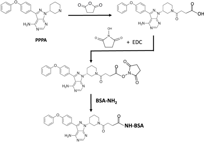
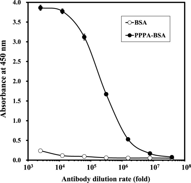
Ibrutinib is an oral irreversible Bruton's tyrosine kinase (BTK) inhibitor that blocks BTK activity by forming covalent bonds with the thiol group of cysteine in the ATP-binding pocket via Michael addition. However, it also reacts with a variety of off-target nonspecific proteins. In this study, we attempted to generate a specific antibody against ibrutinib and develop an immunohistochemical method to detect the ibrutinib-protein conjugates. Ibrutinib has the same amino group as the nucleobase adenine. Paraformaldehyde fixation could not fix it to the tissue via this amino group. However, ibrutinib covalently binds to proteins such as BTKs to exert its action and is therefore immobilized in tissue as ibrutinib-protein conjugates. Thus, immunohistochemistry for ibrutinib detects the location of the ibrutinib-protein conjugates, that is, the sites of covalently bound to the tissue via Michael addition. Using this immunohistochemical method, we visualized the ibrutinib-protein conjugates in the rat gastrointestinal tract (gastric body, duodenum, jejunum, ileum, and colon). This study is the first to elucidate the location of the ibrutinib-protein conjugates in the rat gastrointestinal tract and helps to clarify the mechanism of ibrutinib-induced toxicity.
期刊介绍:
Acta Histochemica et Cytochemica is the official online journal of the Japan Society of Histochemistry and Cytochemistry. It is intended primarily for rapid publication of concise, original articles in the fields of histochemistry and cytochemistry. Manuscripts oriented towards methodological subjects that contain significant technical advances in these fields are also welcome. Manuscripts in English are accepted from investigators in any country, whether or not they are members of the Japan Society of Histochemistry and Cytochemistry. Manuscripts should be original work that has not been previously published and is not being considered for publication elsewhere, with the exception of abstracts. Manuscripts with essentially the same content as a paper that has been published or accepted, or is under consideration for publication, will not be considered. All submitted papers will be peer-reviewed by at least two referees selected by an appropriate Associate Editor. Acceptance is based on scientific significance, originality, and clarity. When required, a revised manuscript should be submitted within 3 months, otherwise it will be considered to be a new submission. The Editor-in-Chief will make all final decisions regarding acceptance.
 求助内容:
求助内容: 应助结果提醒方式:


