Hannah L Weaver, Gabrielle S Fontes, Yi-Fan Shen, Ryan Jennings, Janis M Lapsley, Laura E Selmic
{"title":"狗手术边缘胃肠道肿瘤和正常组织的偏振敏感光学相干断层成像特征。","authors":"Hannah L Weaver, Gabrielle S Fontes, Yi-Fan Shen, Ryan Jennings, Janis M Lapsley, Laura E Selmic","doi":"10.1111/vco.70001","DOIUrl":null,"url":null,"abstract":"<p><p>The treatment of choice for canine alimentary tract neoplasms is surgical excision, but it can sometimes be difficult to achieve wide margins due to neoplasm location, size, or distribution. Optical coherence tomography (OCT) is a rapid, noninvasive imaging modality that uses light to characterise tissue microstructure to allow identification of different tissue types. Spectral domain (SD)-OCT allows for differentiation based on the total light intensity reflected from the tissue. Polarisation sensitive (PS)-OCT detects the polarisation state of light reflected by the tissues. The polarisation properties are phase retardation, degree of polarisation uniformity (DOPU), and optical axis. Our objective was to qualitatively characterise different tissues at the excision sites of alimentary tract neoplasms using OCT. Oral, liver, and other alimentary tumours including stomach, intestine, and pancreas were imaged. Samples were then fixed in formalin, paraffin embedded and stained with haematoxylin and eosin. OCT images and histology slides were compared, and the tissues were qualitatively described by a single investigator. We hypothesized that PS-OCT imaging would provide distinguishing characteristics of tissue appearances that could be used in the future to train observers or artificial intelligence to identify incomplete margins. Our results showed that alimentary tract tumours have disorganised microstructures on SD-OCT and PS-OCT DOPU, and PS-OCT phase retardation and optical axis values that differ from normal tissues. Thus, these characteristics can be used to differentiate neoplastic and normal tissues at surgical margins.</p>","PeriodicalId":23693,"journal":{"name":"Veterinary and comparative oncology","volume":" ","pages":""},"PeriodicalIF":1.9000,"publicationDate":"2025-06-18","publicationTypes":"Journal Article","fieldsOfStudy":null,"isOpenAccess":false,"openAccessPdf":"https://www.ncbi.nlm.nih.gov/pmc/articles/PMC12378641/pdf/","citationCount":"0","resultStr":"{\"title\":\"Polarisation Sensitive Optical Coherence Tomography Image Characteristics for Gastrointestinal Tumours and Normal Tissues at Surgical Margins in Dogs.\",\"authors\":\"Hannah L Weaver, Gabrielle S Fontes, Yi-Fan Shen, Ryan Jennings, Janis M Lapsley, Laura E Selmic\",\"doi\":\"10.1111/vco.70001\",\"DOIUrl\":null,\"url\":null,\"abstract\":\"<p><p>The treatment of choice for canine alimentary tract neoplasms is surgical excision, but it can sometimes be difficult to achieve wide margins due to neoplasm location, size, or distribution. Optical coherence tomography (OCT) is a rapid, noninvasive imaging modality that uses light to characterise tissue microstructure to allow identification of different tissue types. Spectral domain (SD)-OCT allows for differentiation based on the total light intensity reflected from the tissue. Polarisation sensitive (PS)-OCT detects the polarisation state of light reflected by the tissues. The polarisation properties are phase retardation, degree of polarisation uniformity (DOPU), and optical axis. Our objective was to qualitatively characterise different tissues at the excision sites of alimentary tract neoplasms using OCT. Oral, liver, and other alimentary tumours including stomach, intestine, and pancreas were imaged. Samples were then fixed in formalin, paraffin embedded and stained with haematoxylin and eosin. OCT images and histology slides were compared, and the tissues were qualitatively described by a single investigator. We hypothesized that PS-OCT imaging would provide distinguishing characteristics of tissue appearances that could be used in the future to train observers or artificial intelligence to identify incomplete margins. Our results showed that alimentary tract tumours have disorganised microstructures on SD-OCT and PS-OCT DOPU, and PS-OCT phase retardation and optical axis values that differ from normal tissues. Thus, these characteristics can be used to differentiate neoplastic and normal tissues at surgical margins.</p>\",\"PeriodicalId\":23693,\"journal\":{\"name\":\"Veterinary and comparative oncology\",\"volume\":\" \",\"pages\":\"\"},\"PeriodicalIF\":1.9000,\"publicationDate\":\"2025-06-18\",\"publicationTypes\":\"Journal Article\",\"fieldsOfStudy\":null,\"isOpenAccess\":false,\"openAccessPdf\":\"https://www.ncbi.nlm.nih.gov/pmc/articles/PMC12378641/pdf/\",\"citationCount\":\"0\",\"resultStr\":null,\"platform\":\"Semanticscholar\",\"paperid\":null,\"PeriodicalName\":\"Veterinary and comparative oncology\",\"FirstCategoryId\":\"97\",\"ListUrlMain\":\"https://doi.org/10.1111/vco.70001\",\"RegionNum\":2,\"RegionCategory\":\"农林科学\",\"ArticlePicture\":[],\"TitleCN\":null,\"AbstractTextCN\":null,\"PMCID\":null,\"EPubDate\":\"\",\"PubModel\":\"\",\"JCR\":\"Q1\",\"JCRName\":\"VETERINARY SCIENCES\",\"Score\":null,\"Total\":0}","platform":"Semanticscholar","paperid":null,"PeriodicalName":"Veterinary and comparative oncology","FirstCategoryId":"97","ListUrlMain":"https://doi.org/10.1111/vco.70001","RegionNum":2,"RegionCategory":"农林科学","ArticlePicture":[],"TitleCN":null,"AbstractTextCN":null,"PMCID":null,"EPubDate":"","PubModel":"","JCR":"Q1","JCRName":"VETERINARY SCIENCES","Score":null,"Total":0}
Polarisation Sensitive Optical Coherence Tomography Image Characteristics for Gastrointestinal Tumours and Normal Tissues at Surgical Margins in Dogs.
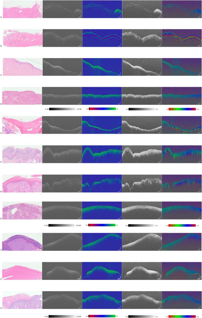
The treatment of choice for canine alimentary tract neoplasms is surgical excision, but it can sometimes be difficult to achieve wide margins due to neoplasm location, size, or distribution. Optical coherence tomography (OCT) is a rapid, noninvasive imaging modality that uses light to characterise tissue microstructure to allow identification of different tissue types. Spectral domain (SD)-OCT allows for differentiation based on the total light intensity reflected from the tissue. Polarisation sensitive (PS)-OCT detects the polarisation state of light reflected by the tissues. The polarisation properties are phase retardation, degree of polarisation uniformity (DOPU), and optical axis. Our objective was to qualitatively characterise different tissues at the excision sites of alimentary tract neoplasms using OCT. Oral, liver, and other alimentary tumours including stomach, intestine, and pancreas were imaged. Samples were then fixed in formalin, paraffin embedded and stained with haematoxylin and eosin. OCT images and histology slides were compared, and the tissues were qualitatively described by a single investigator. We hypothesized that PS-OCT imaging would provide distinguishing characteristics of tissue appearances that could be used in the future to train observers or artificial intelligence to identify incomplete margins. Our results showed that alimentary tract tumours have disorganised microstructures on SD-OCT and PS-OCT DOPU, and PS-OCT phase retardation and optical axis values that differ from normal tissues. Thus, these characteristics can be used to differentiate neoplastic and normal tissues at surgical margins.
期刊介绍:
Veterinary and Comparative Oncology (VCO) is an international, peer-reviewed journal integrating clinical and scientific information from a variety of related disciplines and from worldwide sources for all veterinary oncologists and cancer researchers concerned with aetiology, diagnosis and clinical course of cancer in domestic animals and its prevention. With the ultimate aim of diminishing suffering from cancer, the journal supports the transfer of knowledge in all aspects of veterinary oncology, from the application of new laboratory technology to cancer prevention, early detection, diagnosis and therapy. In addition to original articles, the journal publishes solicited editorials, review articles, commentary, correspondence and abstracts from the published literature. Accordingly, studies describing laboratory work performed exclusively in purpose-bred domestic animals (e.g. dogs, cats, horses) will not be considered.
 求助内容:
求助内容: 应助结果提醒方式:


