Chunlei He, Enyu Yuan, Lei Ye, Hui Xu, Xiaoyong Zhang, Hao Zeng, Yuntian Chen, Jin Yao, Bin Song
{"title":"时间依赖扩散MRI定量分析透明细胞肾细胞癌WHO/ISUP分级","authors":"Chunlei He, Enyu Yuan, Lei Ye, Hui Xu, Xiaoyong Zhang, Hao Zeng, Yuntian Chen, Jin Yao, Bin Song","doi":"10.3348/kjr.2024.1202","DOIUrl":null,"url":null,"abstract":"<p><strong>Objective: </strong>To investigate the feasibility of time-dependent diffusion-weighted imaging (<i>t<sub>d</sub></i>-<i>dMRI</i>) in assessing the pathological World Health Organization/International Society of Urological Pathology (WHO/ISUP) grade of clear cell renal cell carcinoma (ccRCC).</p><p><strong>Materials and methods: </strong>A total of 138 patients (median age, 58 years [interquartile range, 51-64 years]; 89 males) with surgically confirmed ccRCC, comprising 48 high-grade (WHO/ISUP grade III/IV) and 90 low-grade (WHO/ISUP grade I/II) tumors, were included in the study among patients who underwent preoperative <i>t<sub>d</sub></i>-<i>dMRI</i> for suspected RCC between May 2022 and May 2024. The <i>t<sub>d</sub></i>-<i>dMRI</i> microstructural parameters, including cell <i>diameter</i> (<i>d</i>), intracellular volume fraction (<i>f<sub>in</sub></i>), <i>cellularity</i>, and extracellular diffusivities (<i>D<sub>ex</sub></i>), were quantified using a two-compartment model. The solid tumor area was manually annotated to extract the mean values from each parameter map. We analyzed the differences in <i>t<sub>d</sub></i>-<i>dMRI</i> parameters between the high- and low-grade tumors and evaluated the ability of these parameters to distinguish between the two tumor groups. High-definition hematoxylin-and-eosin-stained slides were obtained from 92 patients. We assessed the correlation between <i>t<sub>d</sub></i>-<i>dMRI</i> parameters and pathologic nuclear fraction, which was quantified using an automated nucleus segmentation model (Hover-Net).</p><p><strong>Results: </strong>Compared to high-grade tumors, low-grade tumors exhibited lower <i>cellularity</i> and <i>f<sub>in</sub></i> and higher <i>diameter</i> and <i>D<sub>ex</sub></i>. For differentiation between low- and high-grade ccRCC, the <i>f<sub>in</sub></i> exhibited the highest diagnostic performance (areas under the receiver operating characteristic curve [AUC] = 0.943; 95% confidence interval, 0.906-0.980), followed by <i>cellularity</i> (AUC = 0.931; 0.887-0.976), <i>D<sub>ex</sub></i> (AUC = 0.863; 0.800-0.926), and <i>diameter</i> (AUC = 0.690; 0.596-0.784). The nuclei on pathology slides were automatically segmented, and the nuclear fraction exhibited a moderate correlation with <i>f<sub>in</sub></i> (<i>r</i> = 0.65, <i>P</i> < 0.001).</p><p><strong>Conclusion: </strong><i>t<sub>d</sub></i>-<i>dMRI</i> parameters show potential for assessing pathological WHO/ISUP grades and may serve as promising noninvasive biomarkers for characterizing RCC.</p>","PeriodicalId":17881,"journal":{"name":"Korean Journal of Radiology","volume":" ","pages":"678-687"},"PeriodicalIF":5.3000,"publicationDate":"2025-07-01","publicationTypes":"Journal Article","fieldsOfStudy":null,"isOpenAccess":false,"openAccessPdf":"https://www.ncbi.nlm.nih.gov/pmc/articles/PMC12235544/pdf/","citationCount":"0","resultStr":"{\"title\":\"Quantitative Analysis With Time-Dependent Diffusion MRI for Assessing WHO/ISUP Tumor Grade in Clear Cell Renal Cell Carcinoma.\",\"authors\":\"Chunlei He, Enyu Yuan, Lei Ye, Hui Xu, Xiaoyong Zhang, Hao Zeng, Yuntian Chen, Jin Yao, Bin Song\",\"doi\":\"10.3348/kjr.2024.1202\",\"DOIUrl\":null,\"url\":null,\"abstract\":\"<p><strong>Objective: </strong>To investigate the feasibility of time-dependent diffusion-weighted imaging (<i>t<sub>d</sub></i>-<i>dMRI</i>) in assessing the pathological World Health Organization/International Society of Urological Pathology (WHO/ISUP) grade of clear cell renal cell carcinoma (ccRCC).</p><p><strong>Materials and methods: </strong>A total of 138 patients (median age, 58 years [interquartile range, 51-64 years]; 89 males) with surgically confirmed ccRCC, comprising 48 high-grade (WHO/ISUP grade III/IV) and 90 low-grade (WHO/ISUP grade I/II) tumors, were included in the study among patients who underwent preoperative <i>t<sub>d</sub></i>-<i>dMRI</i> for suspected RCC between May 2022 and May 2024. The <i>t<sub>d</sub></i>-<i>dMRI</i> microstructural parameters, including cell <i>diameter</i> (<i>d</i>), intracellular volume fraction (<i>f<sub>in</sub></i>), <i>cellularity</i>, and extracellular diffusivities (<i>D<sub>ex</sub></i>), were quantified using a two-compartment model. The solid tumor area was manually annotated to extract the mean values from each parameter map. We analyzed the differences in <i>t<sub>d</sub></i>-<i>dMRI</i> parameters between the high- and low-grade tumors and evaluated the ability of these parameters to distinguish between the two tumor groups. High-definition hematoxylin-and-eosin-stained slides were obtained from 92 patients. We assessed the correlation between <i>t<sub>d</sub></i>-<i>dMRI</i> parameters and pathologic nuclear fraction, which was quantified using an automated nucleus segmentation model (Hover-Net).</p><p><strong>Results: </strong>Compared to high-grade tumors, low-grade tumors exhibited lower <i>cellularity</i> and <i>f<sub>in</sub></i> and higher <i>diameter</i> and <i>D<sub>ex</sub></i>. For differentiation between low- and high-grade ccRCC, the <i>f<sub>in</sub></i> exhibited the highest diagnostic performance (areas under the receiver operating characteristic curve [AUC] = 0.943; 95% confidence interval, 0.906-0.980), followed by <i>cellularity</i> (AUC = 0.931; 0.887-0.976), <i>D<sub>ex</sub></i> (AUC = 0.863; 0.800-0.926), and <i>diameter</i> (AUC = 0.690; 0.596-0.784). The nuclei on pathology slides were automatically segmented, and the nuclear fraction exhibited a moderate correlation with <i>f<sub>in</sub></i> (<i>r</i> = 0.65, <i>P</i> < 0.001).</p><p><strong>Conclusion: </strong><i>t<sub>d</sub></i>-<i>dMRI</i> parameters show potential for assessing pathological WHO/ISUP grades and may serve as promising noninvasive biomarkers for characterizing RCC.</p>\",\"PeriodicalId\":17881,\"journal\":{\"name\":\"Korean Journal of Radiology\",\"volume\":\" \",\"pages\":\"678-687\"},\"PeriodicalIF\":5.3000,\"publicationDate\":\"2025-07-01\",\"publicationTypes\":\"Journal Article\",\"fieldsOfStudy\":null,\"isOpenAccess\":false,\"openAccessPdf\":\"https://www.ncbi.nlm.nih.gov/pmc/articles/PMC12235544/pdf/\",\"citationCount\":\"0\",\"resultStr\":null,\"platform\":\"Semanticscholar\",\"paperid\":null,\"PeriodicalName\":\"Korean Journal of Radiology\",\"FirstCategoryId\":\"3\",\"ListUrlMain\":\"https://doi.org/10.3348/kjr.2024.1202\",\"RegionNum\":2,\"RegionCategory\":\"医学\",\"ArticlePicture\":[],\"TitleCN\":null,\"AbstractTextCN\":null,\"PMCID\":null,\"EPubDate\":\"2025/6/13 0:00:00\",\"PubModel\":\"Epub\",\"JCR\":\"Q1\",\"JCRName\":\"RADIOLOGY, NUCLEAR MEDICINE & MEDICAL IMAGING\",\"Score\":null,\"Total\":0}","platform":"Semanticscholar","paperid":null,"PeriodicalName":"Korean Journal of Radiology","FirstCategoryId":"3","ListUrlMain":"https://doi.org/10.3348/kjr.2024.1202","RegionNum":2,"RegionCategory":"医学","ArticlePicture":[],"TitleCN":null,"AbstractTextCN":null,"PMCID":null,"EPubDate":"2025/6/13 0:00:00","PubModel":"Epub","JCR":"Q1","JCRName":"RADIOLOGY, NUCLEAR MEDICINE & MEDICAL IMAGING","Score":null,"Total":0}
Quantitative Analysis With Time-Dependent Diffusion MRI for Assessing WHO/ISUP Tumor Grade in Clear Cell Renal Cell Carcinoma.
Objective: To investigate the feasibility of time-dependent diffusion-weighted imaging (td-dMRI) in assessing the pathological World Health Organization/International Society of Urological Pathology (WHO/ISUP) grade of clear cell renal cell carcinoma (ccRCC).
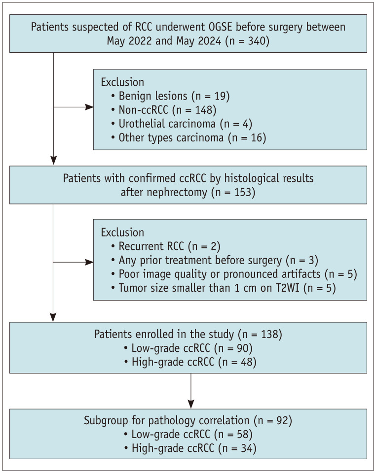
Materials and methods: A total of 138 patients (median age, 58 years [interquartile range, 51-64 years]; 89 males) with surgically confirmed ccRCC, comprising 48 high-grade (WHO/ISUP grade III/IV) and 90 low-grade (WHO/ISUP grade I/II) tumors, were included in the study among patients who underwent preoperative td-dMRI for suspected RCC between May 2022 and May 2024. The td-dMRI microstructural parameters, including cell diameter (d), intracellular volume fraction (fin), cellularity, and extracellular diffusivities (Dex), were quantified using a two-compartment model. The solid tumor area was manually annotated to extract the mean values from each parameter map. We analyzed the differences in td-dMRI parameters between the high- and low-grade tumors and evaluated the ability of these parameters to distinguish between the two tumor groups. High-definition hematoxylin-and-eosin-stained slides were obtained from 92 patients. We assessed the correlation between td-dMRI parameters and pathologic nuclear fraction, which was quantified using an automated nucleus segmentation model (Hover-Net).
Results: Compared to high-grade tumors, low-grade tumors exhibited lower cellularity and fin and higher diameter and Dex. For differentiation between low- and high-grade ccRCC, the fin exhibited the highest diagnostic performance (areas under the receiver operating characteristic curve [AUC] = 0.943; 95% confidence interval, 0.906-0.980), followed by cellularity (AUC = 0.931; 0.887-0.976), Dex (AUC = 0.863; 0.800-0.926), and diameter (AUC = 0.690; 0.596-0.784). The nuclei on pathology slides were automatically segmented, and the nuclear fraction exhibited a moderate correlation with fin (r = 0.65, P < 0.001).
Conclusion: td-dMRI parameters show potential for assessing pathological WHO/ISUP grades and may serve as promising noninvasive biomarkers for characterizing RCC.
期刊介绍:
The inaugural issue of the Korean J Radiol came out in March 2000. Our journal aims to produce and propagate knowledge on radiologic imaging and related sciences.
A unique feature of the articles published in the Journal will be their reflection of global trends in radiology combined with an East-Asian perspective. Geographic differences in disease prevalence will be reflected in the contents of papers, and this will serve to enrich our body of knowledge.
World''s outstanding radiologists from many countries are serving as editorial board of our journal.


 求助内容:
求助内容: 应助结果提醒方式:


