Sunu Mathew, Yen-Ning Huang, Paula Bice, Andrew J Saykin, Shannon L Risacher
{"title":"轻度认知障碍和阿尔茨海默病的视网膜血管生物标志物:一项综合综述和荟萃分析。","authors":"Sunu Mathew, Yen-Ning Huang, Paula Bice, Andrew J Saykin, Shannon L Risacher","doi":"10.1002/dad2.70132","DOIUrl":null,"url":null,"abstract":"<p><p>Retinal vasculature could be a novel, non-invasive, and inexpensive biomarker for Alzheimer's disease (AD) -related neuropathology. In this comprehensive review and meta-analysis from studies that assessed retinal vasculature using optical coherence tomography angiography in AD dementia, we calculated the pooled standardized mean differences (SMDs) in vascular density (VD)-whole, VD-parafoveal, vessel length density (VLD) and foveal avascular zone (FAZ) between AD and cognitively normal (CN), and between mild cognitive impairment (MCI) and CN. Thirty-six studies were included in the meta-analysis. The pooled SMD between AD and CN in VD-whole, VD-parafoveal, VLD, and FAZ was -0.69 (<i>p</i> < 0.01), -0.38 (<i>p</i> = 0.01), -0.59 (<i>p</i> < 0.01), and 0.33 (<i>p</i> = 0.06), respectively, and between MCI and CN in VD-whole, VD-parafoveal, VLD, and FAZ was -0.36 (<i>p</i> = 0.03), -0.17 (<i>p</i> = 0.44), -0.34 (<i>p</i> = 0.03), and 0.31 (<i>p</i> = 0.07), respectively. We identified a significant reduction in retinal vasculature in AD and MCI compared to CN.</p><p><strong>Highlights: </strong>We performed a meta-analysis of 36 studies using optical coherence tomography angiography to assess the retinal vasculature.These included 4129 participants, of which 1175 had Alzheimer's disease (AD), 1004 had mild cognitive impairment, and 1926 were cognitively normal.We identified a significant reduction in retinal vasculature in AD and mild cognitive impairment compared to cognitively normal.Retinal perfusion measured using optical coherence tomography angiography could be used as a potential biomarker for AD dementia.</p>","PeriodicalId":53226,"journal":{"name":"Alzheimer''s and Dementia: Diagnosis, Assessment and Disease Monitoring","volume":"17 2","pages":"e70132"},"PeriodicalIF":4.4000,"publicationDate":"2025-06-16","publicationTypes":"Journal Article","fieldsOfStudy":null,"isOpenAccess":false,"openAccessPdf":"https://www.ncbi.nlm.nih.gov/pmc/articles/PMC12168234/pdf/","citationCount":"0","resultStr":"{\"title\":\"Retinal vascular biomarkers in mild cognitive impairment and Alzheimer's disease: a comprehensive review and meta-analysis.\",\"authors\":\"Sunu Mathew, Yen-Ning Huang, Paula Bice, Andrew J Saykin, Shannon L Risacher\",\"doi\":\"10.1002/dad2.70132\",\"DOIUrl\":null,\"url\":null,\"abstract\":\"<p><p>Retinal vasculature could be a novel, non-invasive, and inexpensive biomarker for Alzheimer's disease (AD) -related neuropathology. In this comprehensive review and meta-analysis from studies that assessed retinal vasculature using optical coherence tomography angiography in AD dementia, we calculated the pooled standardized mean differences (SMDs) in vascular density (VD)-whole, VD-parafoveal, vessel length density (VLD) and foveal avascular zone (FAZ) between AD and cognitively normal (CN), and between mild cognitive impairment (MCI) and CN. Thirty-six studies were included in the meta-analysis. The pooled SMD between AD and CN in VD-whole, VD-parafoveal, VLD, and FAZ was -0.69 (<i>p</i> < 0.01), -0.38 (<i>p</i> = 0.01), -0.59 (<i>p</i> < 0.01), and 0.33 (<i>p</i> = 0.06), respectively, and between MCI and CN in VD-whole, VD-parafoveal, VLD, and FAZ was -0.36 (<i>p</i> = 0.03), -0.17 (<i>p</i> = 0.44), -0.34 (<i>p</i> = 0.03), and 0.31 (<i>p</i> = 0.07), respectively. We identified a significant reduction in retinal vasculature in AD and MCI compared to CN.</p><p><strong>Highlights: </strong>We performed a meta-analysis of 36 studies using optical coherence tomography angiography to assess the retinal vasculature.These included 4129 participants, of which 1175 had Alzheimer's disease (AD), 1004 had mild cognitive impairment, and 1926 were cognitively normal.We identified a significant reduction in retinal vasculature in AD and mild cognitive impairment compared to cognitively normal.Retinal perfusion measured using optical coherence tomography angiography could be used as a potential biomarker for AD dementia.</p>\",\"PeriodicalId\":53226,\"journal\":{\"name\":\"Alzheimer''s and Dementia: Diagnosis, Assessment and Disease Monitoring\",\"volume\":\"17 2\",\"pages\":\"e70132\"},\"PeriodicalIF\":4.4000,\"publicationDate\":\"2025-06-16\",\"publicationTypes\":\"Journal Article\",\"fieldsOfStudy\":null,\"isOpenAccess\":false,\"openAccessPdf\":\"https://www.ncbi.nlm.nih.gov/pmc/articles/PMC12168234/pdf/\",\"citationCount\":\"0\",\"resultStr\":null,\"platform\":\"Semanticscholar\",\"paperid\":null,\"PeriodicalName\":\"Alzheimer''s and Dementia: Diagnosis, Assessment and Disease Monitoring\",\"FirstCategoryId\":\"1085\",\"ListUrlMain\":\"https://doi.org/10.1002/dad2.70132\",\"RegionNum\":0,\"RegionCategory\":null,\"ArticlePicture\":[],\"TitleCN\":null,\"AbstractTextCN\":null,\"PMCID\":null,\"EPubDate\":\"2025/4/1 0:00:00\",\"PubModel\":\"eCollection\",\"JCR\":\"Q1\",\"JCRName\":\"CLINICAL NEUROLOGY\",\"Score\":null,\"Total\":0}","platform":"Semanticscholar","paperid":null,"PeriodicalName":"Alzheimer''s and Dementia: Diagnosis, Assessment and Disease Monitoring","FirstCategoryId":"1085","ListUrlMain":"https://doi.org/10.1002/dad2.70132","RegionNum":0,"RegionCategory":null,"ArticlePicture":[],"TitleCN":null,"AbstractTextCN":null,"PMCID":null,"EPubDate":"2025/4/1 0:00:00","PubModel":"eCollection","JCR":"Q1","JCRName":"CLINICAL NEUROLOGY","Score":null,"Total":0}
Retinal vascular biomarkers in mild cognitive impairment and Alzheimer's disease: a comprehensive review and meta-analysis.
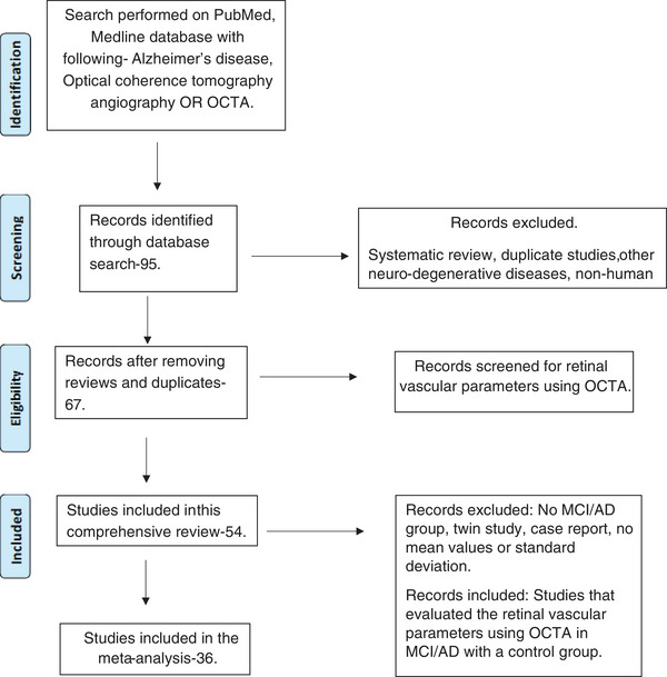
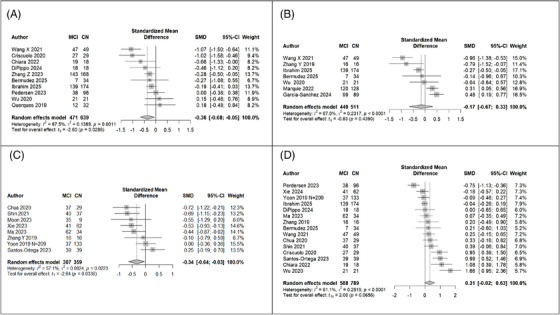
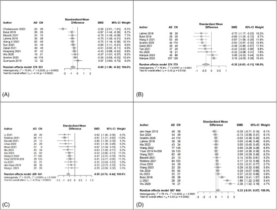
Retinal vasculature could be a novel, non-invasive, and inexpensive biomarker for Alzheimer's disease (AD) -related neuropathology. In this comprehensive review and meta-analysis from studies that assessed retinal vasculature using optical coherence tomography angiography in AD dementia, we calculated the pooled standardized mean differences (SMDs) in vascular density (VD)-whole, VD-parafoveal, vessel length density (VLD) and foveal avascular zone (FAZ) between AD and cognitively normal (CN), and between mild cognitive impairment (MCI) and CN. Thirty-six studies were included in the meta-analysis. The pooled SMD between AD and CN in VD-whole, VD-parafoveal, VLD, and FAZ was -0.69 (p < 0.01), -0.38 (p = 0.01), -0.59 (p < 0.01), and 0.33 (p = 0.06), respectively, and between MCI and CN in VD-whole, VD-parafoveal, VLD, and FAZ was -0.36 (p = 0.03), -0.17 (p = 0.44), -0.34 (p = 0.03), and 0.31 (p = 0.07), respectively. We identified a significant reduction in retinal vasculature in AD and MCI compared to CN.
Highlights: We performed a meta-analysis of 36 studies using optical coherence tomography angiography to assess the retinal vasculature.These included 4129 participants, of which 1175 had Alzheimer's disease (AD), 1004 had mild cognitive impairment, and 1926 were cognitively normal.We identified a significant reduction in retinal vasculature in AD and mild cognitive impairment compared to cognitively normal.Retinal perfusion measured using optical coherence tomography angiography could be used as a potential biomarker for AD dementia.
期刊介绍:
Alzheimer''s & Dementia: Diagnosis, Assessment & Disease Monitoring (DADM) is an open access, peer-reviewed, journal from the Alzheimer''s Association® that will publish new research that reports the discovery, development and validation of instruments, technologies, algorithms, and innovative processes. Papers will cover a range of topics interested in the early and accurate detection of individuals with memory complaints and/or among asymptomatic individuals at elevated risk for various forms of memory disorders. The expectation for published papers will be to translate fundamental knowledge about the neurobiology of the disease into practical reports that describe both the conceptual and methodological aspects of the submitted scientific inquiry. Published topics will explore the development of biomarkers, surrogate markers, and conceptual/methodological challenges. Publication priority will be given to papers that 1) describe putative surrogate markers that accurately track disease progression, 2) biomarkers that fulfill international regulatory requirements, 3) reports from large, well-characterized population-based cohorts that comprise the heterogeneity and diversity of asymptomatic individuals and 4) algorithmic development that considers multi-marker arrays (e.g., integrated-omics, genetics, biofluids, imaging, etc.) and advanced computational analytics and technologies.
 求助内容:
求助内容: 应助结果提醒方式:


