Bo Kyu Choi, Ho Heon Yang, Jong Hyun Kim, JaeSeong Hong, Kyung Min Kim, Yu Rang Park
{"title":"基于脑脊液无标记三维免疫细胞形态学的中枢神经系统感染诊断和预后深度学习模型","authors":"Bo Kyu Choi, Ho Heon Yang, Jong Hyun Kim, JaeSeong Hong, Kyung Min Kim, Yu Rang Park","doi":"10.1002/aisy.202401145","DOIUrl":null,"url":null,"abstract":"<p>Early diagnosis and prognostication of a central nervous system (CNS) infection is essential. This study aims to use immune-cell morphology to develop a deep-learning model for this purpose. Overall, 1427 3D images of cerebrospinal fluid (CSF) immune cells from 14 patients with CNS infections are obtained using holotomography. The images are categorized into infection etiology groups (viral and non-viral) and prognosis groups (based on the modified Rankin Scale score at discharge). A deep-learning model is constructed to predict the etiology and prognosis of CNS infections using the immune-cell morphology. Cell morphological features and spatial distribution of CSF immune cells differ significantly between patients in the viral and nonviral groups and between prognosis groups. The model yields areas under the receiver operating characteristic curve of 0.89 and 0.79 for the diagnosis and prognosis, respectively. As more cell images are used, the prediction and model robustness improve. With <10 cells, both tasks exhibit a nearly 100% predictive performance. After dividing the cells into eight shells, significant refractive index variations are observed. This is the first study to use CSF cell morphology for the diagnosis and prognostication of CSF infections. These findings can help improve patient outcomes.</p>","PeriodicalId":93858,"journal":{"name":"Advanced intelligent systems (Weinheim an der Bergstrasse, Germany)","volume":"7 6","pages":""},"PeriodicalIF":6.1000,"publicationDate":"2025-03-26","publicationTypes":"Journal Article","fieldsOfStudy":null,"isOpenAccess":false,"openAccessPdf":"https://onlinelibrary.wiley.com/doi/epdf/10.1002/aisy.202401145","citationCount":"0","resultStr":"{\"title\":\"Deep-Learning Model for Central Nervous System Infection Diagnosis and Prognosis Using Label-Free 3D Immune-Cell Morphology in the Cerebrospinal Fluid\",\"authors\":\"Bo Kyu Choi, Ho Heon Yang, Jong Hyun Kim, JaeSeong Hong, Kyung Min Kim, Yu Rang Park\",\"doi\":\"10.1002/aisy.202401145\",\"DOIUrl\":null,\"url\":null,\"abstract\":\"<p>Early diagnosis and prognostication of a central nervous system (CNS) infection is essential. This study aims to use immune-cell morphology to develop a deep-learning model for this purpose. Overall, 1427 3D images of cerebrospinal fluid (CSF) immune cells from 14 patients with CNS infections are obtained using holotomography. The images are categorized into infection etiology groups (viral and non-viral) and prognosis groups (based on the modified Rankin Scale score at discharge). A deep-learning model is constructed to predict the etiology and prognosis of CNS infections using the immune-cell morphology. Cell morphological features and spatial distribution of CSF immune cells differ significantly between patients in the viral and nonviral groups and between prognosis groups. The model yields areas under the receiver operating characteristic curve of 0.89 and 0.79 for the diagnosis and prognosis, respectively. As more cell images are used, the prediction and model robustness improve. With <10 cells, both tasks exhibit a nearly 100% predictive performance. After dividing the cells into eight shells, significant refractive index variations are observed. This is the first study to use CSF cell morphology for the diagnosis and prognostication of CSF infections. These findings can help improve patient outcomes.</p>\",\"PeriodicalId\":93858,\"journal\":{\"name\":\"Advanced intelligent systems (Weinheim an der Bergstrasse, Germany)\",\"volume\":\"7 6\",\"pages\":\"\"},\"PeriodicalIF\":6.1000,\"publicationDate\":\"2025-03-26\",\"publicationTypes\":\"Journal Article\",\"fieldsOfStudy\":null,\"isOpenAccess\":false,\"openAccessPdf\":\"https://onlinelibrary.wiley.com/doi/epdf/10.1002/aisy.202401145\",\"citationCount\":\"0\",\"resultStr\":null,\"platform\":\"Semanticscholar\",\"paperid\":null,\"PeriodicalName\":\"Advanced intelligent systems (Weinheim an der Bergstrasse, Germany)\",\"FirstCategoryId\":\"1085\",\"ListUrlMain\":\"https://advanced.onlinelibrary.wiley.com/doi/10.1002/aisy.202401145\",\"RegionNum\":0,\"RegionCategory\":null,\"ArticlePicture\":[],\"TitleCN\":null,\"AbstractTextCN\":null,\"PMCID\":null,\"EPubDate\":\"\",\"PubModel\":\"\",\"JCR\":\"Q1\",\"JCRName\":\"AUTOMATION & CONTROL SYSTEMS\",\"Score\":null,\"Total\":0}","platform":"Semanticscholar","paperid":null,"PeriodicalName":"Advanced intelligent systems (Weinheim an der Bergstrasse, Germany)","FirstCategoryId":"1085","ListUrlMain":"https://advanced.onlinelibrary.wiley.com/doi/10.1002/aisy.202401145","RegionNum":0,"RegionCategory":null,"ArticlePicture":[],"TitleCN":null,"AbstractTextCN":null,"PMCID":null,"EPubDate":"","PubModel":"","JCR":"Q1","JCRName":"AUTOMATION & CONTROL SYSTEMS","Score":null,"Total":0}
Deep-Learning Model for Central Nervous System Infection Diagnosis and Prognosis Using Label-Free 3D Immune-Cell Morphology in the Cerebrospinal Fluid
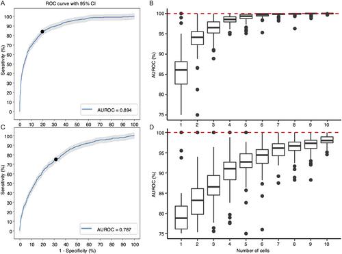
Early diagnosis and prognostication of a central nervous system (CNS) infection is essential. This study aims to use immune-cell morphology to develop a deep-learning model for this purpose. Overall, 1427 3D images of cerebrospinal fluid (CSF) immune cells from 14 patients with CNS infections are obtained using holotomography. The images are categorized into infection etiology groups (viral and non-viral) and prognosis groups (based on the modified Rankin Scale score at discharge). A deep-learning model is constructed to predict the etiology and prognosis of CNS infections using the immune-cell morphology. Cell morphological features and spatial distribution of CSF immune cells differ significantly between patients in the viral and nonviral groups and between prognosis groups. The model yields areas under the receiver operating characteristic curve of 0.89 and 0.79 for the diagnosis and prognosis, respectively. As more cell images are used, the prediction and model robustness improve. With <10 cells, both tasks exhibit a nearly 100% predictive performance. After dividing the cells into eight shells, significant refractive index variations are observed. This is the first study to use CSF cell morphology for the diagnosis and prognostication of CSF infections. These findings can help improve patient outcomes.


 求助内容:
求助内容: 应助结果提醒方式:


