Luay Ibrahim Abu Atileh, Nouf Khalifeh, Caroline Sabanekh, Lina Amairi, Bayan Al Omari
{"title":"带蒂腺肌瘤伴骨化生1例。","authors":"Luay Ibrahim Abu Atileh, Nouf Khalifeh, Caroline Sabanekh, Lina Amairi, Bayan Al Omari","doi":"10.4103/gmit.GMIT-D-24-00026","DOIUrl":null,"url":null,"abstract":"<p><p>We report a case of a pedunculated adenomyoma with osseous metaplasia, which mimics a dermoid cyst on magnetic resonance imaging (MRI) making it a considerable differential diagnosis. A 40-year-old female presented with chronic lower abdomen pain for a year. Pelvic MRI revealed a right-sided pelvic mass measuring 8.4 cm × 5.7 cm. The mass appeared isointense, with hyperintense contents similar to calcifications and fatty content. These results strongly indicated a dermoid cyst. During laparoscopy, a massive pedunculated uterine myoma was seen on the right posterior-fundal part of the uterus. During laparoscopic myomectomy, calcified tissues were discovered during manual morcellation. The histopathological examination confirmed the diagnosis of adenomyoma with widespread calcification and localized osseous metaplasia. Osseous metaplasia is an uncommon cytomorphological transformation seen mostly in the endometrium. Adenomyomas are rare, benign uterine tumors that are frequently misdiagnosed. In this case, the preoperative diagnosis suggested a dermoid cyst, broadening the differential diagnosis for calcified uterine tumors detected on MRI.</p>","PeriodicalId":45272,"journal":{"name":"Gynecology and Minimally Invasive Therapy-GMIT","volume":"14 2","pages":"185-188"},"PeriodicalIF":1.7000,"publicationDate":"2025-05-22","publicationTypes":"Journal Article","fieldsOfStudy":null,"isOpenAccess":false,"openAccessPdf":"https://www.ncbi.nlm.nih.gov/pmc/articles/PMC12165686/pdf/","citationCount":"0","resultStr":"{\"title\":\"Pedunculated Adenomyoma with Osseous Metaplasia: A Case Report.\",\"authors\":\"Luay Ibrahim Abu Atileh, Nouf Khalifeh, Caroline Sabanekh, Lina Amairi, Bayan Al Omari\",\"doi\":\"10.4103/gmit.GMIT-D-24-00026\",\"DOIUrl\":null,\"url\":null,\"abstract\":\"<p><p>We report a case of a pedunculated adenomyoma with osseous metaplasia, which mimics a dermoid cyst on magnetic resonance imaging (MRI) making it a considerable differential diagnosis. A 40-year-old female presented with chronic lower abdomen pain for a year. Pelvic MRI revealed a right-sided pelvic mass measuring 8.4 cm × 5.7 cm. The mass appeared isointense, with hyperintense contents similar to calcifications and fatty content. These results strongly indicated a dermoid cyst. During laparoscopy, a massive pedunculated uterine myoma was seen on the right posterior-fundal part of the uterus. During laparoscopic myomectomy, calcified tissues were discovered during manual morcellation. The histopathological examination confirmed the diagnosis of adenomyoma with widespread calcification and localized osseous metaplasia. Osseous metaplasia is an uncommon cytomorphological transformation seen mostly in the endometrium. Adenomyomas are rare, benign uterine tumors that are frequently misdiagnosed. In this case, the preoperative diagnosis suggested a dermoid cyst, broadening the differential diagnosis for calcified uterine tumors detected on MRI.</p>\",\"PeriodicalId\":45272,\"journal\":{\"name\":\"Gynecology and Minimally Invasive Therapy-GMIT\",\"volume\":\"14 2\",\"pages\":\"185-188\"},\"PeriodicalIF\":1.7000,\"publicationDate\":\"2025-05-22\",\"publicationTypes\":\"Journal Article\",\"fieldsOfStudy\":null,\"isOpenAccess\":false,\"openAccessPdf\":\"https://www.ncbi.nlm.nih.gov/pmc/articles/PMC12165686/pdf/\",\"citationCount\":\"0\",\"resultStr\":null,\"platform\":\"Semanticscholar\",\"paperid\":null,\"PeriodicalName\":\"Gynecology and Minimally Invasive Therapy-GMIT\",\"FirstCategoryId\":\"1085\",\"ListUrlMain\":\"https://doi.org/10.4103/gmit.GMIT-D-24-00026\",\"RegionNum\":0,\"RegionCategory\":null,\"ArticlePicture\":[],\"TitleCN\":null,\"AbstractTextCN\":null,\"PMCID\":null,\"EPubDate\":\"2025/4/1 0:00:00\",\"PubModel\":\"eCollection\",\"JCR\":\"Q3\",\"JCRName\":\"OBSTETRICS & GYNECOLOGY\",\"Score\":null,\"Total\":0}","platform":"Semanticscholar","paperid":null,"PeriodicalName":"Gynecology and Minimally Invasive Therapy-GMIT","FirstCategoryId":"1085","ListUrlMain":"https://doi.org/10.4103/gmit.GMIT-D-24-00026","RegionNum":0,"RegionCategory":null,"ArticlePicture":[],"TitleCN":null,"AbstractTextCN":null,"PMCID":null,"EPubDate":"2025/4/1 0:00:00","PubModel":"eCollection","JCR":"Q3","JCRName":"OBSTETRICS & GYNECOLOGY","Score":null,"Total":0}
引用次数: 0
摘要
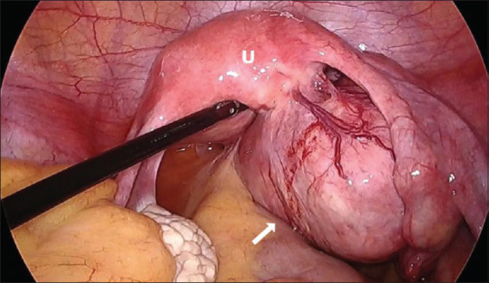
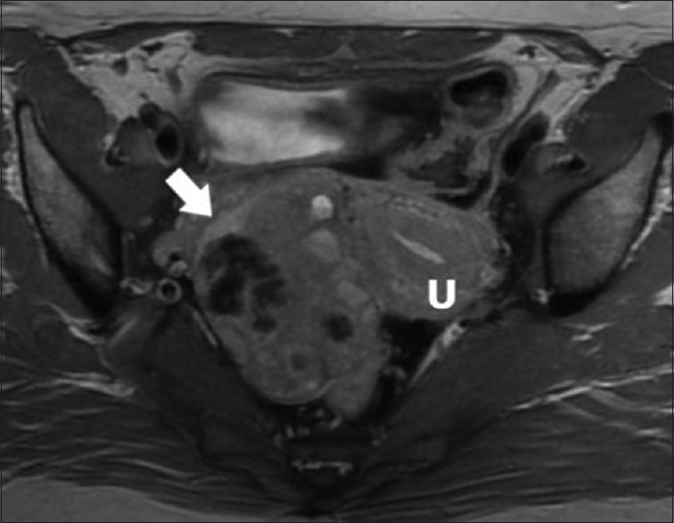
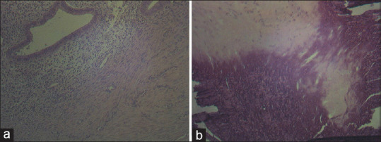
我们报告一例带蒂腺肌瘤伴骨性化生,其在磁共振成像(MRI)上与皮样囊肿相似,使其具有相当的鉴别诊断价值。女性,40岁,慢性下腹部疼痛一年。盆腔MRI示右侧盆腔肿块,大小为8.4 cm × 5.7 cm。肿块呈等强度,高强度的内容物与钙化和脂肪含量相似。这些结果强烈提示为皮样囊肿。腹腔镜检查发现子宫右侧后基底部有一个巨大的带蒂子宫肌瘤。在腹腔镜子宫肌瘤切除术中,在手工分块时发现钙化组织。组织病理学检查证实子宫腺肌瘤伴广泛钙化和局部骨化生。骨化生是一种罕见的细胞形态转变,主要发生在子宫内膜。腺肌瘤是一种罕见的良性子宫肿瘤,常被误诊。本例术前诊断为皮样囊肿,扩大了MRI对钙化子宫肿瘤的鉴别诊断范围。
Pedunculated Adenomyoma with Osseous Metaplasia: A Case Report.
We report a case of a pedunculated adenomyoma with osseous metaplasia, which mimics a dermoid cyst on magnetic resonance imaging (MRI) making it a considerable differential diagnosis. A 40-year-old female presented with chronic lower abdomen pain for a year. Pelvic MRI revealed a right-sided pelvic mass measuring 8.4 cm × 5.7 cm. The mass appeared isointense, with hyperintense contents similar to calcifications and fatty content. These results strongly indicated a dermoid cyst. During laparoscopy, a massive pedunculated uterine myoma was seen on the right posterior-fundal part of the uterus. During laparoscopic myomectomy, calcified tissues were discovered during manual morcellation. The histopathological examination confirmed the diagnosis of adenomyoma with widespread calcification and localized osseous metaplasia. Osseous metaplasia is an uncommon cytomorphological transformation seen mostly in the endometrium. Adenomyomas are rare, benign uterine tumors that are frequently misdiagnosed. In this case, the preoperative diagnosis suggested a dermoid cyst, broadening the differential diagnosis for calcified uterine tumors detected on MRI.

 求助内容:
求助内容: 应助结果提醒方式:


