Ricardo Luz Leitão Guerra, Luiz Roisman, Jay S Duker, Giuseppe Querques, Luiz Filipe Adami Lucatto, Emmerson Badaró, Gabriel Castilho S Barbosa, Eduardo Amorim Novais
{"title":"用常规光谱域光学相干层析成像显示不可见的内丛状层分层。","authors":"Ricardo Luz Leitão Guerra, Luiz Roisman, Jay S Duker, Giuseppe Querques, Luiz Filipe Adami Lucatto, Emmerson Badaró, Gabriel Castilho S Barbosa, Eduardo Amorim Novais","doi":"10.1186/s40942-025-00692-3","DOIUrl":null,"url":null,"abstract":"<p><strong>Background: </strong>The inner plexiform layer (IPL) of the retina plays a key role in visual processing, consisting of five stratified sub-bands (S1-S5) that segregate ON and OFF visual pathways. Until now, resolving these IPL sub-layers was only possible with experimental high-resolution (HR-OCT) or visible-light OCT (VIS-OCT), which remain inaccessible for clinical use. This study provides the first demonstration that IPL stratification can be visualized using commercially available spectral-domain OCT (SD-OCT) with optimized imaging and grayscale inversion.</p><p><strong>Methods: </strong>This retrospective, cross-sectional image analysis study included three healthy individuals who underwent macular OCT imaging. Two subjects were imaged with SD-OCT devices (Nidek RS3000 Advance and Zeiss Cirrus 6000), while one subject was imaged with a swept-source OCT (SS-OCT) device (Topcon Triton DRI). High-density B-scans (1024 A-scans per B-scan) with 120 repetitions for noise reduction were analyzed in both standard and inverted grayscale display modes. The impact of scan size (12 mm, 6 mm, and 3 mm) on IPL visualization was also evaluated.</p><p><strong>Results: </strong>In conventional grayscale, IPL stratification was indistinct. However, inverted grayscale revealed five IPL sub-bands in all cases, particularly in the parafoveal region where the IPL is thicker. Hyperreflective dots near IPL-1, likely representing the superficial capillary plexus, were also identified. The 3-mm scan protocol provided superior sub-layer differentiation compared to 12-mm scans. However, SS-OCT images did not allow for the distinction of the five IPL strata.</p><p><strong>Conclusions: </strong>This study challenges the belief that IPL stratification cannot be identified with conventional SD-OCT. By refining imaging parameters and using grayscale inversion, this approach enhances retinal circuit analysis with standard technology. While SD-OCT enables detailed IPL visualization under specific conditions, SS-OCT does not appear to be well-suited for this purpose. These findings redefine SD-OCT's diagnostic capabilities, opening avenues for research in ophthalmology and neurodegenerative disease monitoring. Further studies should establish best practices and expand clinical applications for this novel methodology.</p>","PeriodicalId":14289,"journal":{"name":"International Journal of Retina and Vitreous","volume":"11 1","pages":"65"},"PeriodicalIF":2.4000,"publicationDate":"2025-06-15","publicationTypes":"Journal Article","fieldsOfStudy":null,"isOpenAccess":false,"openAccessPdf":"https://www.ncbi.nlm.nih.gov/pmc/articles/PMC12168253/pdf/","citationCount":"0","resultStr":"{\"title\":\"Visualizing the invisible: inner plexiform layer stratification with conventional spectral-domain optical coherence tomography.\",\"authors\":\"Ricardo Luz Leitão Guerra, Luiz Roisman, Jay S Duker, Giuseppe Querques, Luiz Filipe Adami Lucatto, Emmerson Badaró, Gabriel Castilho S Barbosa, Eduardo Amorim Novais\",\"doi\":\"10.1186/s40942-025-00692-3\",\"DOIUrl\":null,\"url\":null,\"abstract\":\"<p><strong>Background: </strong>The inner plexiform layer (IPL) of the retina plays a key role in visual processing, consisting of five stratified sub-bands (S1-S5) that segregate ON and OFF visual pathways. Until now, resolving these IPL sub-layers was only possible with experimental high-resolution (HR-OCT) or visible-light OCT (VIS-OCT), which remain inaccessible for clinical use. This study provides the first demonstration that IPL stratification can be visualized using commercially available spectral-domain OCT (SD-OCT) with optimized imaging and grayscale inversion.</p><p><strong>Methods: </strong>This retrospective, cross-sectional image analysis study included three healthy individuals who underwent macular OCT imaging. Two subjects were imaged with SD-OCT devices (Nidek RS3000 Advance and Zeiss Cirrus 6000), while one subject was imaged with a swept-source OCT (SS-OCT) device (Topcon Triton DRI). High-density B-scans (1024 A-scans per B-scan) with 120 repetitions for noise reduction were analyzed in both standard and inverted grayscale display modes. The impact of scan size (12 mm, 6 mm, and 3 mm) on IPL visualization was also evaluated.</p><p><strong>Results: </strong>In conventional grayscale, IPL stratification was indistinct. However, inverted grayscale revealed five IPL sub-bands in all cases, particularly in the parafoveal region where the IPL is thicker. Hyperreflective dots near IPL-1, likely representing the superficial capillary plexus, were also identified. The 3-mm scan protocol provided superior sub-layer differentiation compared to 12-mm scans. However, SS-OCT images did not allow for the distinction of the five IPL strata.</p><p><strong>Conclusions: </strong>This study challenges the belief that IPL stratification cannot be identified with conventional SD-OCT. By refining imaging parameters and using grayscale inversion, this approach enhances retinal circuit analysis with standard technology. While SD-OCT enables detailed IPL visualization under specific conditions, SS-OCT does not appear to be well-suited for this purpose. These findings redefine SD-OCT's diagnostic capabilities, opening avenues for research in ophthalmology and neurodegenerative disease monitoring. Further studies should establish best practices and expand clinical applications for this novel methodology.</p>\",\"PeriodicalId\":14289,\"journal\":{\"name\":\"International Journal of Retina and Vitreous\",\"volume\":\"11 1\",\"pages\":\"65\"},\"PeriodicalIF\":2.4000,\"publicationDate\":\"2025-06-15\",\"publicationTypes\":\"Journal Article\",\"fieldsOfStudy\":null,\"isOpenAccess\":false,\"openAccessPdf\":\"https://www.ncbi.nlm.nih.gov/pmc/articles/PMC12168253/pdf/\",\"citationCount\":\"0\",\"resultStr\":null,\"platform\":\"Semanticscholar\",\"paperid\":null,\"PeriodicalName\":\"International Journal of Retina and Vitreous\",\"FirstCategoryId\":\"1085\",\"ListUrlMain\":\"https://doi.org/10.1186/s40942-025-00692-3\",\"RegionNum\":0,\"RegionCategory\":null,\"ArticlePicture\":[],\"TitleCN\":null,\"AbstractTextCN\":null,\"PMCID\":null,\"EPubDate\":\"\",\"PubModel\":\"\",\"JCR\":\"Q2\",\"JCRName\":\"OPHTHALMOLOGY\",\"Score\":null,\"Total\":0}","platform":"Semanticscholar","paperid":null,"PeriodicalName":"International Journal of Retina and Vitreous","FirstCategoryId":"1085","ListUrlMain":"https://doi.org/10.1186/s40942-025-00692-3","RegionNum":0,"RegionCategory":null,"ArticlePicture":[],"TitleCN":null,"AbstractTextCN":null,"PMCID":null,"EPubDate":"","PubModel":"","JCR":"Q2","JCRName":"OPHTHALMOLOGY","Score":null,"Total":0}
Visualizing the invisible: inner plexiform layer stratification with conventional spectral-domain optical coherence tomography.
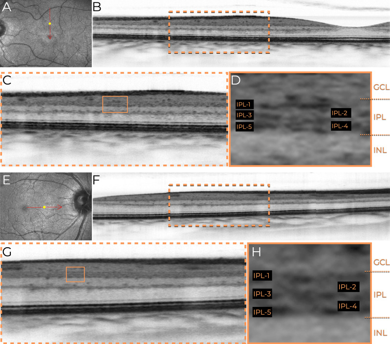
Background: The inner plexiform layer (IPL) of the retina plays a key role in visual processing, consisting of five stratified sub-bands (S1-S5) that segregate ON and OFF visual pathways. Until now, resolving these IPL sub-layers was only possible with experimental high-resolution (HR-OCT) or visible-light OCT (VIS-OCT), which remain inaccessible for clinical use. This study provides the first demonstration that IPL stratification can be visualized using commercially available spectral-domain OCT (SD-OCT) with optimized imaging and grayscale inversion.
Methods: This retrospective, cross-sectional image analysis study included three healthy individuals who underwent macular OCT imaging. Two subjects were imaged with SD-OCT devices (Nidek RS3000 Advance and Zeiss Cirrus 6000), while one subject was imaged with a swept-source OCT (SS-OCT) device (Topcon Triton DRI). High-density B-scans (1024 A-scans per B-scan) with 120 repetitions for noise reduction were analyzed in both standard and inverted grayscale display modes. The impact of scan size (12 mm, 6 mm, and 3 mm) on IPL visualization was also evaluated.
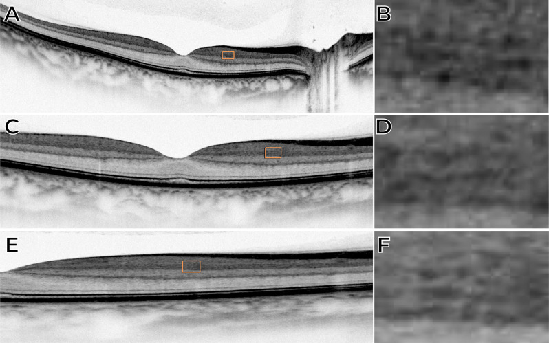
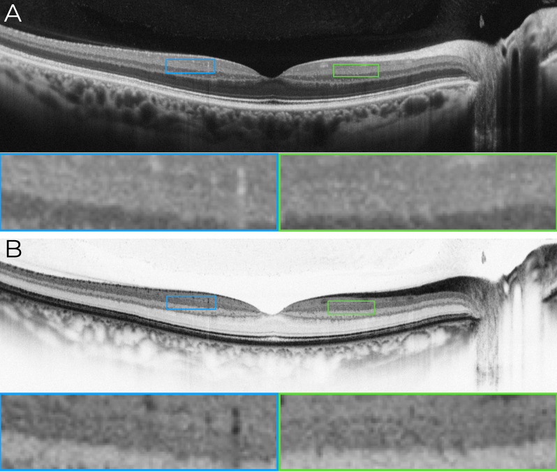
Results: In conventional grayscale, IPL stratification was indistinct. However, inverted grayscale revealed five IPL sub-bands in all cases, particularly in the parafoveal region where the IPL is thicker. Hyperreflective dots near IPL-1, likely representing the superficial capillary plexus, were also identified. The 3-mm scan protocol provided superior sub-layer differentiation compared to 12-mm scans. However, SS-OCT images did not allow for the distinction of the five IPL strata.
Conclusions: This study challenges the belief that IPL stratification cannot be identified with conventional SD-OCT. By refining imaging parameters and using grayscale inversion, this approach enhances retinal circuit analysis with standard technology. While SD-OCT enables detailed IPL visualization under specific conditions, SS-OCT does not appear to be well-suited for this purpose. These findings redefine SD-OCT's diagnostic capabilities, opening avenues for research in ophthalmology and neurodegenerative disease monitoring. Further studies should establish best practices and expand clinical applications for this novel methodology.
期刊介绍:
International Journal of Retina and Vitreous focuses on the ophthalmic subspecialty of vitreoretinal disorders. The journal presents original articles on new approaches to diagnosis, outcomes of clinical trials, innovations in pharmacological therapy and surgical techniques, as well as basic science advances that impact clinical practice. Topical areas include, but are not limited to: -Imaging of the retina, choroid and vitreous -Innovations in optical coherence tomography (OCT) -Small-gauge vitrectomy, retinal detachment, chromovitrectomy -Electroretinography (ERG), microperimetry, other functional tests -Intraocular tumors -Retinal pharmacotherapy & drug delivery -Diabetic retinopathy & other vascular diseases -Age-related macular degeneration (AMD) & other macular entities

 求助内容:
求助内容: 应助结果提醒方式:


