Oliver B Dilger, Mason F Carstens, Daphne J Garrett, Daniel J Berry, Joaquin Sanchez-Sotelo, Mark E Morrey, Roman Thaler, Amel Dudakovic, Matthew P Abdel
{"title":"转化生长因子-β1 (TGF-β1)在原代人膝关节成纤维细胞中的时间和浓度依赖性作用","authors":"Oliver B Dilger, Mason F Carstens, Daphne J Garrett, Daniel J Berry, Joaquin Sanchez-Sotelo, Mark E Morrey, Roman Thaler, Amel Dudakovic, Matthew P Abdel","doi":"10.1302/2046-3758.146.BJR-2024-0387.R3","DOIUrl":null,"url":null,"abstract":"<p><strong>Aims: </strong>To characterize the effects of varying experimental parameters of transforming growth factor-beta 1 (TGF-β1) in primary knee fibroblasts as an in vitro model of arthrofibrosis.</p><p><strong>Methods: </strong>Primary knee fibroblasts were isolated from one patient undergoing primary total knee arthroplasty (TKA) and one patient undergoing revision TKA for arthrofibrosis, respectively. Fibroblasts were stimulated with varying concentrations and durations of the pro-fibrotic cytokine TGF-β1. Picrosirius red staining (PSR) was conducted to assess collagen deposition, and real-time quantitative polymerase chain reaction (RT-qPCR) and western blotting were performed to determine cellular gene expression levels. A collagen gel contraction assay was used to assess TGF-β1-induced fibroblast contraction.</p><p><strong>Results: </strong>In our experiments, TGF-β1-mediated collagen deposition was consistent across a vast concentration range (0.5 to 40 ng/ml). While α-smooth muscle actin (ACTA2) protein levels and SMAD2 phosphorylation were induced by low concentrations (1 ng/ml), robust <i>ACTA2</i> transcription required higher concentrations (5 ng/ml) of TGF-β1. Evaluation of TGF-β1-mediated cell and cytoskeletal contractility shows that in the measured fibroblasts, this process occurs within three hours following TGF-β1 administration. Of note, TGF-β1-differentiated myofibroblasts exhibited greater contractile properties than naïve fibroblasts in the presence of TGF-β1.</p><p><strong>Conclusion: </strong>Taken together, this study elucidates key TGF-β1 experimental parameters that will inform the development and interpretation of in vitro models of arthrofibrosis.</p>","PeriodicalId":9074,"journal":{"name":"Bone & Joint Research","volume":"14 6","pages":"527-538"},"PeriodicalIF":5.1000,"publicationDate":"2025-06-16","publicationTypes":"Journal Article","fieldsOfStudy":null,"isOpenAccess":false,"openAccessPdf":"https://www.ncbi.nlm.nih.gov/pmc/articles/PMC12168093/pdf/","citationCount":"0","resultStr":"{\"title\":\"Temporal- and concentration-dependent effects of transforming growth factor-beta 1 (TGF-β1) in primary human knee fibroblasts.\",\"authors\":\"Oliver B Dilger, Mason F Carstens, Daphne J Garrett, Daniel J Berry, Joaquin Sanchez-Sotelo, Mark E Morrey, Roman Thaler, Amel Dudakovic, Matthew P Abdel\",\"doi\":\"10.1302/2046-3758.146.BJR-2024-0387.R3\",\"DOIUrl\":null,\"url\":null,\"abstract\":\"<p><strong>Aims: </strong>To characterize the effects of varying experimental parameters of transforming growth factor-beta 1 (TGF-β1) in primary knee fibroblasts as an in vitro model of arthrofibrosis.</p><p><strong>Methods: </strong>Primary knee fibroblasts were isolated from one patient undergoing primary total knee arthroplasty (TKA) and one patient undergoing revision TKA for arthrofibrosis, respectively. Fibroblasts were stimulated with varying concentrations and durations of the pro-fibrotic cytokine TGF-β1. Picrosirius red staining (PSR) was conducted to assess collagen deposition, and real-time quantitative polymerase chain reaction (RT-qPCR) and western blotting were performed to determine cellular gene expression levels. A collagen gel contraction assay was used to assess TGF-β1-induced fibroblast contraction.</p><p><strong>Results: </strong>In our experiments, TGF-β1-mediated collagen deposition was consistent across a vast concentration range (0.5 to 40 ng/ml). While α-smooth muscle actin (ACTA2) protein levels and SMAD2 phosphorylation were induced by low concentrations (1 ng/ml), robust <i>ACTA2</i> transcription required higher concentrations (5 ng/ml) of TGF-β1. Evaluation of TGF-β1-mediated cell and cytoskeletal contractility shows that in the measured fibroblasts, this process occurs within three hours following TGF-β1 administration. Of note, TGF-β1-differentiated myofibroblasts exhibited greater contractile properties than naïve fibroblasts in the presence of TGF-β1.</p><p><strong>Conclusion: </strong>Taken together, this study elucidates key TGF-β1 experimental parameters that will inform the development and interpretation of in vitro models of arthrofibrosis.</p>\",\"PeriodicalId\":9074,\"journal\":{\"name\":\"Bone & Joint Research\",\"volume\":\"14 6\",\"pages\":\"527-538\"},\"PeriodicalIF\":5.1000,\"publicationDate\":\"2025-06-16\",\"publicationTypes\":\"Journal Article\",\"fieldsOfStudy\":null,\"isOpenAccess\":false,\"openAccessPdf\":\"https://www.ncbi.nlm.nih.gov/pmc/articles/PMC12168093/pdf/\",\"citationCount\":\"0\",\"resultStr\":null,\"platform\":\"Semanticscholar\",\"paperid\":null,\"PeriodicalName\":\"Bone & Joint Research\",\"FirstCategoryId\":\"3\",\"ListUrlMain\":\"https://doi.org/10.1302/2046-3758.146.BJR-2024-0387.R3\",\"RegionNum\":2,\"RegionCategory\":\"医学\",\"ArticlePicture\":[],\"TitleCN\":null,\"AbstractTextCN\":null,\"PMCID\":null,\"EPubDate\":\"\",\"PubModel\":\"\",\"JCR\":\"Q2\",\"JCRName\":\"CELL & TISSUE ENGINEERING\",\"Score\":null,\"Total\":0}","platform":"Semanticscholar","paperid":null,"PeriodicalName":"Bone & Joint Research","FirstCategoryId":"3","ListUrlMain":"https://doi.org/10.1302/2046-3758.146.BJR-2024-0387.R3","RegionNum":2,"RegionCategory":"医学","ArticlePicture":[],"TitleCN":null,"AbstractTextCN":null,"PMCID":null,"EPubDate":"","PubModel":"","JCR":"Q2","JCRName":"CELL & TISSUE ENGINEERING","Score":null,"Total":0}
Temporal- and concentration-dependent effects of transforming growth factor-beta 1 (TGF-β1) in primary human knee fibroblasts.
Aims: To characterize the effects of varying experimental parameters of transforming growth factor-beta 1 (TGF-β1) in primary knee fibroblasts as an in vitro model of arthrofibrosis.
Methods: Primary knee fibroblasts were isolated from one patient undergoing primary total knee arthroplasty (TKA) and one patient undergoing revision TKA for arthrofibrosis, respectively. Fibroblasts were stimulated with varying concentrations and durations of the pro-fibrotic cytokine TGF-β1. Picrosirius red staining (PSR) was conducted to assess collagen deposition, and real-time quantitative polymerase chain reaction (RT-qPCR) and western blotting were performed to determine cellular gene expression levels. A collagen gel contraction assay was used to assess TGF-β1-induced fibroblast contraction.
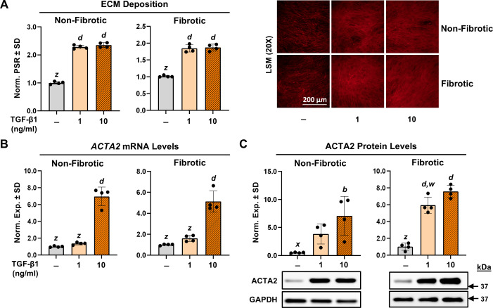
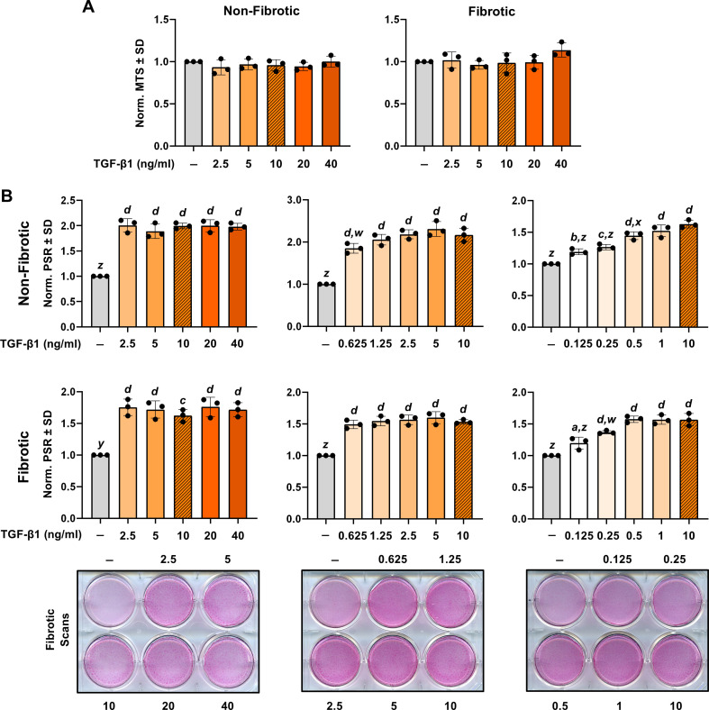
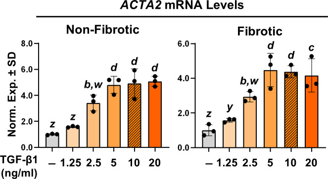
Results: In our experiments, TGF-β1-mediated collagen deposition was consistent across a vast concentration range (0.5 to 40 ng/ml). While α-smooth muscle actin (ACTA2) protein levels and SMAD2 phosphorylation were induced by low concentrations (1 ng/ml), robust ACTA2 transcription required higher concentrations (5 ng/ml) of TGF-β1. Evaluation of TGF-β1-mediated cell and cytoskeletal contractility shows that in the measured fibroblasts, this process occurs within three hours following TGF-β1 administration. Of note, TGF-β1-differentiated myofibroblasts exhibited greater contractile properties than naïve fibroblasts in the presence of TGF-β1.
Conclusion: Taken together, this study elucidates key TGF-β1 experimental parameters that will inform the development and interpretation of in vitro models of arthrofibrosis.
 求助内容:
求助内容: 应助结果提醒方式:


