Emiliano Marasco, Christina Düsing, Stefanie Keymel, Alessandra Bortoluzzi, Claudia Bracaglia, Matthieu Canuet, Ilaria Cavazzana, Gamal Chehab, Veronica Codullo, Rebecca Fischer, Franco Franceschini, Micaela Fredi, Stefano Ghio, Lisa Keller, Alain Meyer, Carlomaurizio Montecucco, Jutta Richter, Marianne Riou, Sezgin Sahin, Oliver Sander, Andreas Schwarting, Carlo Alberto Scirè, Ettore Silvagni, Konstantinos Triantafyllias, Giovanni Zanframundo, Lorenzo Cavagna, Matthias Schneider
{"title":"系统性红斑狼疮肺动脉高压:多中心回顾性队列中危险因素和血流动力学特征的识别。","authors":"Emiliano Marasco, Christina Düsing, Stefanie Keymel, Alessandra Bortoluzzi, Claudia Bracaglia, Matthieu Canuet, Ilaria Cavazzana, Gamal Chehab, Veronica Codullo, Rebecca Fischer, Franco Franceschini, Micaela Fredi, Stefano Ghio, Lisa Keller, Alain Meyer, Carlomaurizio Montecucco, Jutta Richter, Marianne Riou, Sezgin Sahin, Oliver Sander, Andreas Schwarting, Carlo Alberto Scirè, Ettore Silvagni, Konstantinos Triantafyllias, Giovanni Zanframundo, Lorenzo Cavagna, Matthias Schneider","doi":"10.1136/lupus-2024-001471","DOIUrl":null,"url":null,"abstract":"<p><strong>Objectives: </strong>The aim of our work was to identify specific patterns in clinical features and nailfold capillary changes that may help in screening for pulmonary arterial hypertension (PAH) in patients with systemic lupus erythematosus (SLE).</p><p><strong>Methods: </strong>We identified patients with SLE and type I PAH (n=20) without other connective tissue diseases and collected demographic, clinical and laboratory features. We selected as controls patients with SLE who underwent cardiopulmonary screening to exclude PAH (n=87): we collected demographic, clinical and laboratory features and performed nailfold videocapillaroscopy (NVC).</p><p><strong>Results: </strong>All patients with SLE-PAH were women; age and disease duration were not different from patients with SLE without PAH. Lupus anticoagulant (LAC)+and anti-ribonucleoprotein (RNP)+were more prevalent in patients with SLE-PAH (respectively, PAH 45.0% vs no-PAH 20.5%, p=0.042; PAH 45.0% vs no-PAH 19.5%, p=0.035). No differences were observed for anti-Sm, anti-Ro, anti-La and anti-cardiolipin and anti-beta2GPI antibodies. Among clinical features, mucocutaneous and central nervous system involvement were more prevalent in patients with SLE-PAH than in SLE controls (respectively, PAH 65.0% vs no-PAH 34.5%, p=0.024; PAH 25.0% vs no-PAH 8.0%, p=0.046). Raynaud's phenomenon (RP) was more prevalent in patients with SLE-PAH than in SLE controls (PAH 60.0% vs no-PAH 13.8%, p<0.001). RP was a predictor of PAH in patients with SLE (OR 3.8 (0.9-14.8)). We performed NVC on nine patients with PAH and on controls: we observed a significantly higher prevalence of scleroderma pattern at NVC in SLE-PAH than controls (PAH 66.7% vs no-PAH 9.2%, p<0.001). Patients with SLE-PAH showed a lower number of capillary density and a higher frequency of giant capillaries.</p><p><strong>Conclusions: </strong>Our data showed that LAC+, RNP+, RP and a scleroderma pattern at NVC was indicative for patients with SLE-PAH. Our results pointed to generalised microvascular involvement and a hypercoagulation state in patients with SLE-PAH. The variables we identified could be used to implement a screening algorithm to identify patients with SLE at risk of developing PAH.</p>","PeriodicalId":18126,"journal":{"name":"Lupus Science & Medicine","volume":"12 1","pages":""},"PeriodicalIF":3.5000,"publicationDate":"2025-06-12","publicationTypes":"Journal Article","fieldsOfStudy":null,"isOpenAccess":false,"openAccessPdf":"https://www.ncbi.nlm.nih.gov/pmc/articles/PMC12164622/pdf/","citationCount":"0","resultStr":"{\"title\":\"Pulmonary arterial hypertension in systemic lupus erythematosus: identification of risk factors and haemodynamics characteristics in a multicentre retrospective cohort.\",\"authors\":\"Emiliano Marasco, Christina Düsing, Stefanie Keymel, Alessandra Bortoluzzi, Claudia Bracaglia, Matthieu Canuet, Ilaria Cavazzana, Gamal Chehab, Veronica Codullo, Rebecca Fischer, Franco Franceschini, Micaela Fredi, Stefano Ghio, Lisa Keller, Alain Meyer, Carlomaurizio Montecucco, Jutta Richter, Marianne Riou, Sezgin Sahin, Oliver Sander, Andreas Schwarting, Carlo Alberto Scirè, Ettore Silvagni, Konstantinos Triantafyllias, Giovanni Zanframundo, Lorenzo Cavagna, Matthias Schneider\",\"doi\":\"10.1136/lupus-2024-001471\",\"DOIUrl\":null,\"url\":null,\"abstract\":\"<p><strong>Objectives: </strong>The aim of our work was to identify specific patterns in clinical features and nailfold capillary changes that may help in screening for pulmonary arterial hypertension (PAH) in patients with systemic lupus erythematosus (SLE).</p><p><strong>Methods: </strong>We identified patients with SLE and type I PAH (n=20) without other connective tissue diseases and collected demographic, clinical and laboratory features. We selected as controls patients with SLE who underwent cardiopulmonary screening to exclude PAH (n=87): we collected demographic, clinical and laboratory features and performed nailfold videocapillaroscopy (NVC).</p><p><strong>Results: </strong>All patients with SLE-PAH were women; age and disease duration were not different from patients with SLE without PAH. Lupus anticoagulant (LAC)+and anti-ribonucleoprotein (RNP)+were more prevalent in patients with SLE-PAH (respectively, PAH 45.0% vs no-PAH 20.5%, p=0.042; PAH 45.0% vs no-PAH 19.5%, p=0.035). No differences were observed for anti-Sm, anti-Ro, anti-La and anti-cardiolipin and anti-beta2GPI antibodies. Among clinical features, mucocutaneous and central nervous system involvement were more prevalent in patients with SLE-PAH than in SLE controls (respectively, PAH 65.0% vs no-PAH 34.5%, p=0.024; PAH 25.0% vs no-PAH 8.0%, p=0.046). Raynaud's phenomenon (RP) was more prevalent in patients with SLE-PAH than in SLE controls (PAH 60.0% vs no-PAH 13.8%, p<0.001). RP was a predictor of PAH in patients with SLE (OR 3.8 (0.9-14.8)). We performed NVC on nine patients with PAH and on controls: we observed a significantly higher prevalence of scleroderma pattern at NVC in SLE-PAH than controls (PAH 66.7% vs no-PAH 9.2%, p<0.001). Patients with SLE-PAH showed a lower number of capillary density and a higher frequency of giant capillaries.</p><p><strong>Conclusions: </strong>Our data showed that LAC+, RNP+, RP and a scleroderma pattern at NVC was indicative for patients with SLE-PAH. Our results pointed to generalised microvascular involvement and a hypercoagulation state in patients with SLE-PAH. The variables we identified could be used to implement a screening algorithm to identify patients with SLE at risk of developing PAH.</p>\",\"PeriodicalId\":18126,\"journal\":{\"name\":\"Lupus Science & Medicine\",\"volume\":\"12 1\",\"pages\":\"\"},\"PeriodicalIF\":3.5000,\"publicationDate\":\"2025-06-12\",\"publicationTypes\":\"Journal Article\",\"fieldsOfStudy\":null,\"isOpenAccess\":false,\"openAccessPdf\":\"https://www.ncbi.nlm.nih.gov/pmc/articles/PMC12164622/pdf/\",\"citationCount\":\"0\",\"resultStr\":null,\"platform\":\"Semanticscholar\",\"paperid\":null,\"PeriodicalName\":\"Lupus Science & Medicine\",\"FirstCategoryId\":\"3\",\"ListUrlMain\":\"https://doi.org/10.1136/lupus-2024-001471\",\"RegionNum\":2,\"RegionCategory\":\"医学\",\"ArticlePicture\":[],\"TitleCN\":null,\"AbstractTextCN\":null,\"PMCID\":null,\"EPubDate\":\"\",\"PubModel\":\"\",\"JCR\":\"Q1\",\"JCRName\":\"RHEUMATOLOGY\",\"Score\":null,\"Total\":0}","platform":"Semanticscholar","paperid":null,"PeriodicalName":"Lupus Science & Medicine","FirstCategoryId":"3","ListUrlMain":"https://doi.org/10.1136/lupus-2024-001471","RegionNum":2,"RegionCategory":"医学","ArticlePicture":[],"TitleCN":null,"AbstractTextCN":null,"PMCID":null,"EPubDate":"","PubModel":"","JCR":"Q1","JCRName":"RHEUMATOLOGY","Score":null,"Total":0}
Pulmonary arterial hypertension in systemic lupus erythematosus: identification of risk factors and haemodynamics characteristics in a multicentre retrospective cohort.
Objectives: The aim of our work was to identify specific patterns in clinical features and nailfold capillary changes that may help in screening for pulmonary arterial hypertension (PAH) in patients with systemic lupus erythematosus (SLE).
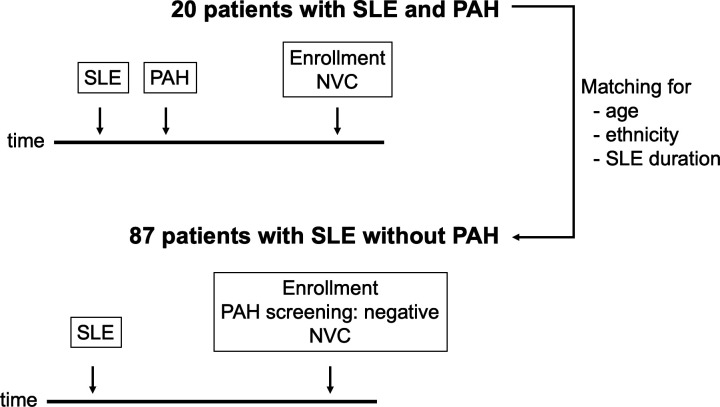
Methods: We identified patients with SLE and type I PAH (n=20) without other connective tissue diseases and collected demographic, clinical and laboratory features. We selected as controls patients with SLE who underwent cardiopulmonary screening to exclude PAH (n=87): we collected demographic, clinical and laboratory features and performed nailfold videocapillaroscopy (NVC).
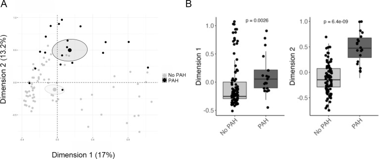
Results: All patients with SLE-PAH were women; age and disease duration were not different from patients with SLE without PAH. Lupus anticoagulant (LAC)+and anti-ribonucleoprotein (RNP)+were more prevalent in patients with SLE-PAH (respectively, PAH 45.0% vs no-PAH 20.5%, p=0.042; PAH 45.0% vs no-PAH 19.5%, p=0.035). No differences were observed for anti-Sm, anti-Ro, anti-La and anti-cardiolipin and anti-beta2GPI antibodies. Among clinical features, mucocutaneous and central nervous system involvement were more prevalent in patients with SLE-PAH than in SLE controls (respectively, PAH 65.0% vs no-PAH 34.5%, p=0.024; PAH 25.0% vs no-PAH 8.0%, p=0.046). Raynaud's phenomenon (RP) was more prevalent in patients with SLE-PAH than in SLE controls (PAH 60.0% vs no-PAH 13.8%, p<0.001). RP was a predictor of PAH in patients with SLE (OR 3.8 (0.9-14.8)). We performed NVC on nine patients with PAH and on controls: we observed a significantly higher prevalence of scleroderma pattern at NVC in SLE-PAH than controls (PAH 66.7% vs no-PAH 9.2%, p<0.001). Patients with SLE-PAH showed a lower number of capillary density and a higher frequency of giant capillaries.
Conclusions: Our data showed that LAC+, RNP+, RP and a scleroderma pattern at NVC was indicative for patients with SLE-PAH. Our results pointed to generalised microvascular involvement and a hypercoagulation state in patients with SLE-PAH. The variables we identified could be used to implement a screening algorithm to identify patients with SLE at risk of developing PAH.
期刊介绍:
Lupus Science & Medicine is a global, peer reviewed, open access online journal that provides a central point for publication of basic, clinical, translational, and epidemiological studies of all aspects of lupus and related diseases. It is the first lupus-specific open access journal in the world and was developed in response to the need for a barrier-free forum for publication of groundbreaking studies in lupus. The journal publishes research on lupus from fields including, but not limited to: rheumatology, dermatology, nephrology, immunology, pediatrics, cardiology, hepatology, pulmonology, obstetrics and gynecology, and psychiatry.
 求助内容:
求助内容: 应助结果提醒方式:


