{"title":"口腔梭形细胞血管瘤的免疫组化分析。","authors":"Amit Mani, Manas Bajpai, Saurabh L Sabnis","doi":"10.30476/dentjods.2024.101499.2305","DOIUrl":null,"url":null,"abstract":"<p><p>Spindle cell hemangioma (SCH), formerly called \"spindle cell hemangioendothelioma\", is a rare benign histological variant of hemangioma characterized by the presence of two contrast zones, the first zone exhibits large dilated cavernous space with slit-like vascular spaces may show clear endothelial vacuoles resembling fat cells. SCH is often considered as pseudosarcomatous entity; it imposes a diagnostic challenge for oral pathologists due to its resemblance with Kaposi sarcoma. A total of 13 cases of SCH have been reported in the head and neck region to date and only 6 cases have been reported inside the oral cavity. We present a rare case of SCH located on the hard palate, which imitated Kaposi's sarcoma on histopathological examination. The expressions of various markers including EGR, CD 31, and HHV 8 yielded the final diagnosis of SCH. The markers EGR and HHV 8 have never been used in intraoral SCH before to the best of our knowledge; hence, the present report highlights the use of immunohistochemistry for the diagnosis of SCH.</p>","PeriodicalId":73702,"journal":{"name":"Journal of dentistry (Shiraz, Iran)","volume":"26 2","pages":"194-198"},"PeriodicalIF":0.0000,"publicationDate":"2025-06-01","publicationTypes":"Journal Article","fieldsOfStudy":null,"isOpenAccess":false,"openAccessPdf":"https://www.ncbi.nlm.nih.gov/pmc/articles/PMC12153497/pdf/","citationCount":"0","resultStr":"{\"title\":\"Immunohistochemical Analysis of Oral Spindle Cell Hemangioma.\",\"authors\":\"Amit Mani, Manas Bajpai, Saurabh L Sabnis\",\"doi\":\"10.30476/dentjods.2024.101499.2305\",\"DOIUrl\":null,\"url\":null,\"abstract\":\"<p><p>Spindle cell hemangioma (SCH), formerly called \\\"spindle cell hemangioendothelioma\\\", is a rare benign histological variant of hemangioma characterized by the presence of two contrast zones, the first zone exhibits large dilated cavernous space with slit-like vascular spaces may show clear endothelial vacuoles resembling fat cells. SCH is often considered as pseudosarcomatous entity; it imposes a diagnostic challenge for oral pathologists due to its resemblance with Kaposi sarcoma. A total of 13 cases of SCH have been reported in the head and neck region to date and only 6 cases have been reported inside the oral cavity. We present a rare case of SCH located on the hard palate, which imitated Kaposi's sarcoma on histopathological examination. The expressions of various markers including EGR, CD 31, and HHV 8 yielded the final diagnosis of SCH. The markers EGR and HHV 8 have never been used in intraoral SCH before to the best of our knowledge; hence, the present report highlights the use of immunohistochemistry for the diagnosis of SCH.</p>\",\"PeriodicalId\":73702,\"journal\":{\"name\":\"Journal of dentistry (Shiraz, Iran)\",\"volume\":\"26 2\",\"pages\":\"194-198\"},\"PeriodicalIF\":0.0000,\"publicationDate\":\"2025-06-01\",\"publicationTypes\":\"Journal Article\",\"fieldsOfStudy\":null,\"isOpenAccess\":false,\"openAccessPdf\":\"https://www.ncbi.nlm.nih.gov/pmc/articles/PMC12153497/pdf/\",\"citationCount\":\"0\",\"resultStr\":null,\"platform\":\"Semanticscholar\",\"paperid\":null,\"PeriodicalName\":\"Journal of dentistry (Shiraz, Iran)\",\"FirstCategoryId\":\"1085\",\"ListUrlMain\":\"https://doi.org/10.30476/dentjods.2024.101499.2305\",\"RegionNum\":0,\"RegionCategory\":null,\"ArticlePicture\":[],\"TitleCN\":null,\"AbstractTextCN\":null,\"PMCID\":null,\"EPubDate\":\"\",\"PubModel\":\"\",\"JCR\":\"\",\"JCRName\":\"\",\"Score\":null,\"Total\":0}","platform":"Semanticscholar","paperid":null,"PeriodicalName":"Journal of dentistry (Shiraz, Iran)","FirstCategoryId":"1085","ListUrlMain":"https://doi.org/10.30476/dentjods.2024.101499.2305","RegionNum":0,"RegionCategory":null,"ArticlePicture":[],"TitleCN":null,"AbstractTextCN":null,"PMCID":null,"EPubDate":"","PubModel":"","JCR":"","JCRName":"","Score":null,"Total":0}
Immunohistochemical Analysis of Oral Spindle Cell Hemangioma.
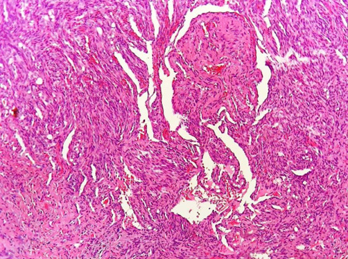
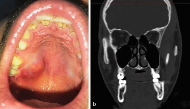
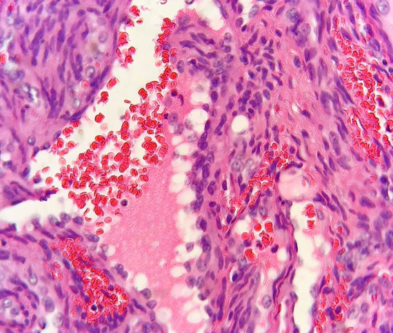
Spindle cell hemangioma (SCH), formerly called "spindle cell hemangioendothelioma", is a rare benign histological variant of hemangioma characterized by the presence of two contrast zones, the first zone exhibits large dilated cavernous space with slit-like vascular spaces may show clear endothelial vacuoles resembling fat cells. SCH is often considered as pseudosarcomatous entity; it imposes a diagnostic challenge for oral pathologists due to its resemblance with Kaposi sarcoma. A total of 13 cases of SCH have been reported in the head and neck region to date and only 6 cases have been reported inside the oral cavity. We present a rare case of SCH located on the hard palate, which imitated Kaposi's sarcoma on histopathological examination. The expressions of various markers including EGR, CD 31, and HHV 8 yielded the final diagnosis of SCH. The markers EGR and HHV 8 have never been used in intraoral SCH before to the best of our knowledge; hence, the present report highlights the use of immunohistochemistry for the diagnosis of SCH.

 求助内容:
求助内容: 应助结果提醒方式:


