Fatemeh Sanie-Jahromi, Abtin Khosravi, Hooman Hadianfard, M Hossein Nowroozzadeh
{"title":"常规胰岛素、葡氨酸胰岛素和天门肽胰岛素对高血糖视网膜色素上皮细胞(RPE)和人视网膜内皮细胞(HRECs)血管内皮生长因子和血管紧张素原表达的影响。","authors":"Fatemeh Sanie-Jahromi, Abtin Khosravi, Hooman Hadianfard, M Hossein Nowroozzadeh","doi":"10.3389/fopht.2025.1570232","DOIUrl":null,"url":null,"abstract":"<p><strong>Introduction: </strong>Diabetic retinopathy (DR) is a leading cause of vision loss and is primarily driven by chronic hyperglycemia, which induces retinal vascular damage through mechanisms involving vascular endothelial growth factor (VEGF) and the renin-angiotensin system (RAS). This study investigated the effects of hyperglycemia and different insulin formulations-regular, glulisine, and aspart-on VEGF-A and angiotensinogen (AGT) gene expression in two human retinal cell types: retinal pigment epithelial (RPE) cells and human retinal microvascular endothelial cells (HRECs).</p><p><strong>Methods: </strong>Cells were cultured from donor tissue and exposed to physiologic and hyperglycemic glucose concentrations, with or without insulin treatment. Gene expression levels were quantified using real-time PCR.</p><p><strong>Results: </strong>Hyperglycemia significantly upregulated VEGF-A and AGT in both RPE and HREC cells (e.g., VEGF-A in RPE: 2.62-fold, <i>P</i> = 0.001; AGT in RPE: 3.32-fold, <i>P</i> = 0.093), supporting a role for both osmotic and glucose-specific pathways. Among insulin treatments, regular insulin significantly reduced VEGF-A expression in both RPE (0.72-fold, <i>P</i> = 0.033) and HRECs (0.57-fold, <i>P</i> = 0.009). In contrast, aspart and glulisine had modest effects on VEGF-A in HRECs (0.82-fold each; <i>P</i> = 0.035 and <i>P</i> = 0.060, respectively) and no significant impact in RPE cells. Regarding AGT, aspart insulin showed the most consistent suppressive effect, reducing expression in both RPE (0.15-fold, <i>P</i> < 0.001) and HRECs (0.22-fold, <i>P</i> = 0.004). Glulisine significantly increased AGT in RPE (1.56-fold, <i>P</i> = 0.009) but reduced it in HRECs (0.58-fold, <i>P</i> = 0.074). Regular insulin showed no effect on AGT in RPE (<i>P</i> = 0.680) and a non-significant increase in HRECs (1.36-fold, <i>P</i> = 0.097).</p><p><strong>Discussion: </strong>These findings highlight the differential biological effects of insulin analogues and suggest that aspart insulin, in particular, may offer therapeutic benefits beyond glycemic control by modulating both VEGF-A and RAS-related pathways. Tailored insulin therapies could represent innovative strategies for managing or slowing the progression of diabetic retinopathy.</p>","PeriodicalId":73096,"journal":{"name":"Frontiers in ophthalmology","volume":"5 ","pages":"1570232"},"PeriodicalIF":0.9000,"publicationDate":"2025-05-29","publicationTypes":"Journal Article","fieldsOfStudy":null,"isOpenAccess":false,"openAccessPdf":"https://www.ncbi.nlm.nih.gov/pmc/articles/PMC12158669/pdf/","citationCount":"0","resultStr":"{\"title\":\"Effects of regular, glulisine, and aspart insulin on vascular endothelial growth factor and angiotensinogen expression in hyperglycemic retinal pigment epithelial (RPE) and human retinal endothelial cells (HRECs).\",\"authors\":\"Fatemeh Sanie-Jahromi, Abtin Khosravi, Hooman Hadianfard, M Hossein Nowroozzadeh\",\"doi\":\"10.3389/fopht.2025.1570232\",\"DOIUrl\":null,\"url\":null,\"abstract\":\"<p><strong>Introduction: </strong>Diabetic retinopathy (DR) is a leading cause of vision loss and is primarily driven by chronic hyperglycemia, which induces retinal vascular damage through mechanisms involving vascular endothelial growth factor (VEGF) and the renin-angiotensin system (RAS). This study investigated the effects of hyperglycemia and different insulin formulations-regular, glulisine, and aspart-on VEGF-A and angiotensinogen (AGT) gene expression in two human retinal cell types: retinal pigment epithelial (RPE) cells and human retinal microvascular endothelial cells (HRECs).</p><p><strong>Methods: </strong>Cells were cultured from donor tissue and exposed to physiologic and hyperglycemic glucose concentrations, with or without insulin treatment. Gene expression levels were quantified using real-time PCR.</p><p><strong>Results: </strong>Hyperglycemia significantly upregulated VEGF-A and AGT in both RPE and HREC cells (e.g., VEGF-A in RPE: 2.62-fold, <i>P</i> = 0.001; AGT in RPE: 3.32-fold, <i>P</i> = 0.093), supporting a role for both osmotic and glucose-specific pathways. Among insulin treatments, regular insulin significantly reduced VEGF-A expression in both RPE (0.72-fold, <i>P</i> = 0.033) and HRECs (0.57-fold, <i>P</i> = 0.009). In contrast, aspart and glulisine had modest effects on VEGF-A in HRECs (0.82-fold each; <i>P</i> = 0.035 and <i>P</i> = 0.060, respectively) and no significant impact in RPE cells. Regarding AGT, aspart insulin showed the most consistent suppressive effect, reducing expression in both RPE (0.15-fold, <i>P</i> < 0.001) and HRECs (0.22-fold, <i>P</i> = 0.004). Glulisine significantly increased AGT in RPE (1.56-fold, <i>P</i> = 0.009) but reduced it in HRECs (0.58-fold, <i>P</i> = 0.074). Regular insulin showed no effect on AGT in RPE (<i>P</i> = 0.680) and a non-significant increase in HRECs (1.36-fold, <i>P</i> = 0.097).</p><p><strong>Discussion: </strong>These findings highlight the differential biological effects of insulin analogues and suggest that aspart insulin, in particular, may offer therapeutic benefits beyond glycemic control by modulating both VEGF-A and RAS-related pathways. Tailored insulin therapies could represent innovative strategies for managing or slowing the progression of diabetic retinopathy.</p>\",\"PeriodicalId\":73096,\"journal\":{\"name\":\"Frontiers in ophthalmology\",\"volume\":\"5 \",\"pages\":\"1570232\"},\"PeriodicalIF\":0.9000,\"publicationDate\":\"2025-05-29\",\"publicationTypes\":\"Journal Article\",\"fieldsOfStudy\":null,\"isOpenAccess\":false,\"openAccessPdf\":\"https://www.ncbi.nlm.nih.gov/pmc/articles/PMC12158669/pdf/\",\"citationCount\":\"0\",\"resultStr\":null,\"platform\":\"Semanticscholar\",\"paperid\":null,\"PeriodicalName\":\"Frontiers in ophthalmology\",\"FirstCategoryId\":\"1085\",\"ListUrlMain\":\"https://doi.org/10.3389/fopht.2025.1570232\",\"RegionNum\":0,\"RegionCategory\":null,\"ArticlePicture\":[],\"TitleCN\":null,\"AbstractTextCN\":null,\"PMCID\":null,\"EPubDate\":\"2025/1/1 0:00:00\",\"PubModel\":\"eCollection\",\"JCR\":\"\",\"JCRName\":\"\",\"Score\":null,\"Total\":0}","platform":"Semanticscholar","paperid":null,"PeriodicalName":"Frontiers in ophthalmology","FirstCategoryId":"1085","ListUrlMain":"https://doi.org/10.3389/fopht.2025.1570232","RegionNum":0,"RegionCategory":null,"ArticlePicture":[],"TitleCN":null,"AbstractTextCN":null,"PMCID":null,"EPubDate":"2025/1/1 0:00:00","PubModel":"eCollection","JCR":"","JCRName":"","Score":null,"Total":0}
Effects of regular, glulisine, and aspart insulin on vascular endothelial growth factor and angiotensinogen expression in hyperglycemic retinal pigment epithelial (RPE) and human retinal endothelial cells (HRECs).
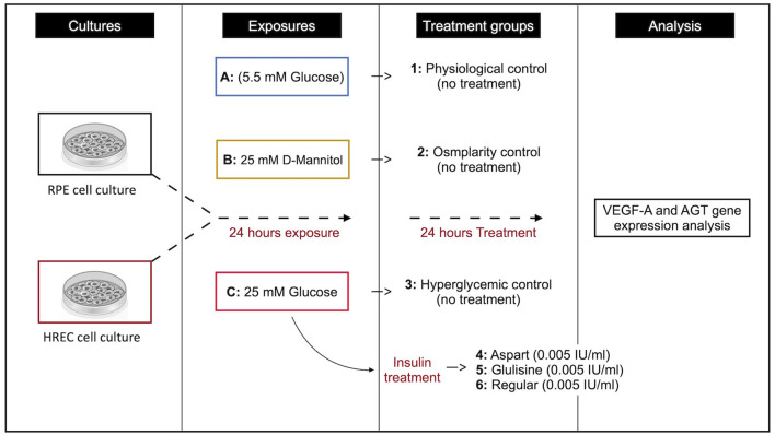
Introduction: Diabetic retinopathy (DR) is a leading cause of vision loss and is primarily driven by chronic hyperglycemia, which induces retinal vascular damage through mechanisms involving vascular endothelial growth factor (VEGF) and the renin-angiotensin system (RAS). This study investigated the effects of hyperglycemia and different insulin formulations-regular, glulisine, and aspart-on VEGF-A and angiotensinogen (AGT) gene expression in two human retinal cell types: retinal pigment epithelial (RPE) cells and human retinal microvascular endothelial cells (HRECs).
Methods: Cells were cultured from donor tissue and exposed to physiologic and hyperglycemic glucose concentrations, with or without insulin treatment. Gene expression levels were quantified using real-time PCR.
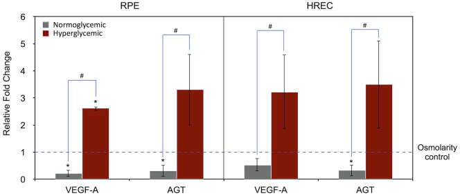
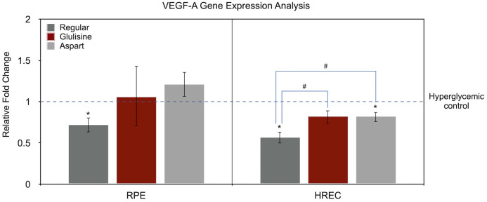
Results: Hyperglycemia significantly upregulated VEGF-A and AGT in both RPE and HREC cells (e.g., VEGF-A in RPE: 2.62-fold, P = 0.001; AGT in RPE: 3.32-fold, P = 0.093), supporting a role for both osmotic and glucose-specific pathways. Among insulin treatments, regular insulin significantly reduced VEGF-A expression in both RPE (0.72-fold, P = 0.033) and HRECs (0.57-fold, P = 0.009). In contrast, aspart and glulisine had modest effects on VEGF-A in HRECs (0.82-fold each; P = 0.035 and P = 0.060, respectively) and no significant impact in RPE cells. Regarding AGT, aspart insulin showed the most consistent suppressive effect, reducing expression in both RPE (0.15-fold, P < 0.001) and HRECs (0.22-fold, P = 0.004). Glulisine significantly increased AGT in RPE (1.56-fold, P = 0.009) but reduced it in HRECs (0.58-fold, P = 0.074). Regular insulin showed no effect on AGT in RPE (P = 0.680) and a non-significant increase in HRECs (1.36-fold, P = 0.097).
Discussion: These findings highlight the differential biological effects of insulin analogues and suggest that aspart insulin, in particular, may offer therapeutic benefits beyond glycemic control by modulating both VEGF-A and RAS-related pathways. Tailored insulin therapies could represent innovative strategies for managing or slowing the progression of diabetic retinopathy.

 求助内容:
求助内容: 应助结果提醒方式:


