Sevdalina Kandilarova, Eleonora Maggioni, Letizia Squarcina, Diyana Najar, Maysam Homadi, Emma Tassi, Drozdstoy Stoyanov, Paolo Brambilla
{"title":"情绪障碍的多变量脑形态模式:额颞叶和小脑区域的关键作用。","authors":"Sevdalina Kandilarova, Eleonora Maggioni, Letizia Squarcina, Diyana Najar, Maysam Homadi, Emma Tassi, Drozdstoy Stoyanov, Paolo Brambilla","doi":"10.1136/bmjment-2024-301511","DOIUrl":null,"url":null,"abstract":"<p><strong>Background: </strong>Differentiating major depressive disorder (MDD) from bipolar disorder (BD) remains a significant clinical challenge, as both disorders exhibit overlapping symptoms but require distinct treatment approaches. Advances in voxel-based morphometry and surface-based morphometry have facilitated the identification of structural brain abnormalities that may serve as diagnostic biomarkers.</p><p><strong>Objective: </strong>This study aimed to explore the relationships between brain morphological features, such as grey matter volume (GMV) and cortical thickness (CT), and demographic and clinical variables in patients with MDD and BD and healthy controls (HC) using multivariate analysis methods.</p><p><strong>Methods: </strong>A total of 263 participants, including 120 HC, 95 patients with MDD and 48 patients with BD, underwent T1-weighted MRI. GMV and CT were computed for standardised brain regions, followed by multivariate partial least squares (PLS) regression to assess associations with demographic and diagnostic variables.</p><p><strong>Findings: </strong>Reductions in frontotemporal CT were observed in MDD and BD compared with HC, but distinct trends between BD and MDD were also detected for the CT of selective temporal, frontal and parietal regions. Differential patterns in cerebellar GMV were also identified, with lobule CI larger in MDD and lobule CII larger in BD. Additionally, BD showed the same trend as ageing concerning reductions in CT and posterior cerebellar and striatal GMV. Depression severity showed a transdiagnostic link with reduced frontotemporal CT.</p><p><strong>Conclusions: </strong>This study highlights shared and distinct structural brain alterations in MDD and BD, emphasising the potential of neuroimaging biomarkers to enhance diagnostic accuracy. Accelerated cortical thinning and differential cerebellar changes in BD may serve as targets for future research and clinical interventions.</p><p><strong>Clinical implications: </strong>Our findings underscore the value of objective neuroimaging markers in increasing the precision of mood disorder diagnoses, improving treatment outcomes.</p>","PeriodicalId":72434,"journal":{"name":"BMJ mental health","volume":"28 1","pages":""},"PeriodicalIF":4.9000,"publicationDate":"2025-06-10","publicationTypes":"Journal Article","fieldsOfStudy":null,"isOpenAccess":false,"openAccessPdf":"https://www.ncbi.nlm.nih.gov/pmc/articles/PMC12161407/pdf/","citationCount":"0","resultStr":"{\"title\":\"Multivariate brain morphological patterns across mood disorders: key roles of frontotemporal and cerebellar areas.\",\"authors\":\"Sevdalina Kandilarova, Eleonora Maggioni, Letizia Squarcina, Diyana Najar, Maysam Homadi, Emma Tassi, Drozdstoy Stoyanov, Paolo Brambilla\",\"doi\":\"10.1136/bmjment-2024-301511\",\"DOIUrl\":null,\"url\":null,\"abstract\":\"<p><strong>Background: </strong>Differentiating major depressive disorder (MDD) from bipolar disorder (BD) remains a significant clinical challenge, as both disorders exhibit overlapping symptoms but require distinct treatment approaches. Advances in voxel-based morphometry and surface-based morphometry have facilitated the identification of structural brain abnormalities that may serve as diagnostic biomarkers.</p><p><strong>Objective: </strong>This study aimed to explore the relationships between brain morphological features, such as grey matter volume (GMV) and cortical thickness (CT), and demographic and clinical variables in patients with MDD and BD and healthy controls (HC) using multivariate analysis methods.</p><p><strong>Methods: </strong>A total of 263 participants, including 120 HC, 95 patients with MDD and 48 patients with BD, underwent T1-weighted MRI. GMV and CT were computed for standardised brain regions, followed by multivariate partial least squares (PLS) regression to assess associations with demographic and diagnostic variables.</p><p><strong>Findings: </strong>Reductions in frontotemporal CT were observed in MDD and BD compared with HC, but distinct trends between BD and MDD were also detected for the CT of selective temporal, frontal and parietal regions. Differential patterns in cerebellar GMV were also identified, with lobule CI larger in MDD and lobule CII larger in BD. Additionally, BD showed the same trend as ageing concerning reductions in CT and posterior cerebellar and striatal GMV. Depression severity showed a transdiagnostic link with reduced frontotemporal CT.</p><p><strong>Conclusions: </strong>This study highlights shared and distinct structural brain alterations in MDD and BD, emphasising the potential of neuroimaging biomarkers to enhance diagnostic accuracy. Accelerated cortical thinning and differential cerebellar changes in BD may serve as targets for future research and clinical interventions.</p><p><strong>Clinical implications: </strong>Our findings underscore the value of objective neuroimaging markers in increasing the precision of mood disorder diagnoses, improving treatment outcomes.</p>\",\"PeriodicalId\":72434,\"journal\":{\"name\":\"BMJ mental health\",\"volume\":\"28 1\",\"pages\":\"\"},\"PeriodicalIF\":4.9000,\"publicationDate\":\"2025-06-10\",\"publicationTypes\":\"Journal Article\",\"fieldsOfStudy\":null,\"isOpenAccess\":false,\"openAccessPdf\":\"https://www.ncbi.nlm.nih.gov/pmc/articles/PMC12161407/pdf/\",\"citationCount\":\"0\",\"resultStr\":null,\"platform\":\"Semanticscholar\",\"paperid\":null,\"PeriodicalName\":\"BMJ mental health\",\"FirstCategoryId\":\"1085\",\"ListUrlMain\":\"https://doi.org/10.1136/bmjment-2024-301511\",\"RegionNum\":0,\"RegionCategory\":null,\"ArticlePicture\":[],\"TitleCN\":null,\"AbstractTextCN\":null,\"PMCID\":null,\"EPubDate\":\"\",\"PubModel\":\"\",\"JCR\":\"0\",\"JCRName\":\"PSYCHIATRY\",\"Score\":null,\"Total\":0}","platform":"Semanticscholar","paperid":null,"PeriodicalName":"BMJ mental health","FirstCategoryId":"1085","ListUrlMain":"https://doi.org/10.1136/bmjment-2024-301511","RegionNum":0,"RegionCategory":null,"ArticlePicture":[],"TitleCN":null,"AbstractTextCN":null,"PMCID":null,"EPubDate":"","PubModel":"","JCR":"0","JCRName":"PSYCHIATRY","Score":null,"Total":0}
Multivariate brain morphological patterns across mood disorders: key roles of frontotemporal and cerebellar areas.
Background: Differentiating major depressive disorder (MDD) from bipolar disorder (BD) remains a significant clinical challenge, as both disorders exhibit overlapping symptoms but require distinct treatment approaches. Advances in voxel-based morphometry and surface-based morphometry have facilitated the identification of structural brain abnormalities that may serve as diagnostic biomarkers.
Objective: This study aimed to explore the relationships between brain morphological features, such as grey matter volume (GMV) and cortical thickness (CT), and demographic and clinical variables in patients with MDD and BD and healthy controls (HC) using multivariate analysis methods.
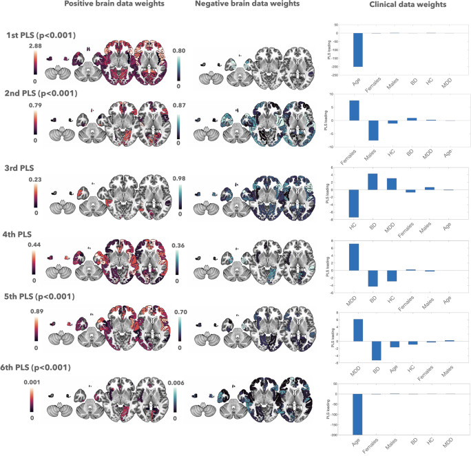
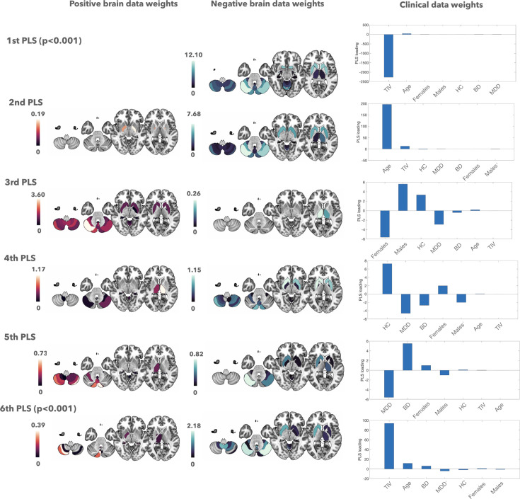
Methods: A total of 263 participants, including 120 HC, 95 patients with MDD and 48 patients with BD, underwent T1-weighted MRI. GMV and CT were computed for standardised brain regions, followed by multivariate partial least squares (PLS) regression to assess associations with demographic and diagnostic variables.
Findings: Reductions in frontotemporal CT were observed in MDD and BD compared with HC, but distinct trends between BD and MDD were also detected for the CT of selective temporal, frontal and parietal regions. Differential patterns in cerebellar GMV were also identified, with lobule CI larger in MDD and lobule CII larger in BD. Additionally, BD showed the same trend as ageing concerning reductions in CT and posterior cerebellar and striatal GMV. Depression severity showed a transdiagnostic link with reduced frontotemporal CT.
Conclusions: This study highlights shared and distinct structural brain alterations in MDD and BD, emphasising the potential of neuroimaging biomarkers to enhance diagnostic accuracy. Accelerated cortical thinning and differential cerebellar changes in BD may serve as targets for future research and clinical interventions.
Clinical implications: Our findings underscore the value of objective neuroimaging markers in increasing the precision of mood disorder diagnoses, improving treatment outcomes.

 求助内容:
求助内容: 应助结果提醒方式:


