Jia-Liang Chen, Shao-Jie Duan, Sheng Xie, Shu-Kun Yao
{"title":"磁共振成像质子密度脂肪分数作为金标准诊断无创脂肪变性生物标志物的准确性。","authors":"Jia-Liang Chen, Shao-Jie Duan, Sheng Xie, Shu-Kun Yao","doi":"10.4329/wjr.v17.i5.104272","DOIUrl":null,"url":null,"abstract":"<p><strong>Background: </strong>Nonalcoholic fatty liver disease (NAFLD) is the most common chronic liver disease. The accuracy of noninvasive biomarkers for detecting hepatic steatosis is still limited.</p><p><strong>Aim: </strong>To assess the diagnostic performance of noninvasive steatosis biomarkers in diagnosing NAFLD using magnetic resonance imaging proton density fat fraction (MRI-PDFF) as the gold standard.</p><p><strong>Methods: </strong>A total of 131 suspected NAFLD patients (60% male, median age 36 years) undergoing MRI-PDFF were consecutively recruited from a tertiary hospital. Steatosis grades determined by MRI-PDFF were classified as none (< 5%), mild (5%-11%), moderate (11%-17%), and severe (≥ 17%). Six steatosis biomarkers were calculated according to clinical parameters and laboratory tests, including fatty liver index, hepatic steatosis index, ZJU index, Framingham steatosis index, triglycerides and glucose index, and visceral adiposity index. The accuracy of these biomarkers in detecting hepatic steatosis was evaluated using the area under the receiver operating characteristic curves (AUCs). The Youden index was used to determine the optimal cut-off for each biomarker. The linear trend analysis of each biomarker across the steatosis grades was conducted by Mantel-Haenszel <i>χ</i> <sup>2</sup> test. Spearman's rank correlation assessed the relationship between steatosis biomarkers and MRI-PDFF.</p><p><strong>Results: </strong>Steatosis grades based on MRI-PDFF prevalence were: None 27%, mild 40%, moderate 15% and severe 18%. Six steatosis biomarkers showed a linear trend across the steatosis grades and a significant positive correlation with MRI-PDFF. The six steatosis biomarkers demonstrated AUCs near 0.90 (range: 0.857-0.912, all <i>P</i> < 0.001) for diagnosing NAFLD by MRI-PDFF ≥ 5%. The optimal cut-offs showed sensitivity between 84.4%-91.7% and specificity between 71.4%-85.7%. The diagnostic performance of these biomarkers in detecting moderate-to-severe and severe steatosis was relatively weaker.</p><p><strong>Conclusion: </strong>These noninvasive steatosis biomarkers accurately diagnosed NAFLD and correlated well with MRI-PDFF for detecting NAFLD, but they did not effectively detect moderate or severe steatosis.</p>","PeriodicalId":23819,"journal":{"name":"World journal of radiology","volume":"17 5","pages":"104272"},"PeriodicalIF":1.5000,"publicationDate":"2025-05-28","publicationTypes":"Journal Article","fieldsOfStudy":null,"isOpenAccess":false,"openAccessPdf":"https://www.ncbi.nlm.nih.gov/pmc/articles/PMC12149971/pdf/","citationCount":"0","resultStr":"{\"title\":\"Diagnostic accuracy of noninvasive steatosis biomarkers with magnetic resonance imaging proton density fat fraction as gold standard.\",\"authors\":\"Jia-Liang Chen, Shao-Jie Duan, Sheng Xie, Shu-Kun Yao\",\"doi\":\"10.4329/wjr.v17.i5.104272\",\"DOIUrl\":null,\"url\":null,\"abstract\":\"<p><strong>Background: </strong>Nonalcoholic fatty liver disease (NAFLD) is the most common chronic liver disease. The accuracy of noninvasive biomarkers for detecting hepatic steatosis is still limited.</p><p><strong>Aim: </strong>To assess the diagnostic performance of noninvasive steatosis biomarkers in diagnosing NAFLD using magnetic resonance imaging proton density fat fraction (MRI-PDFF) as the gold standard.</p><p><strong>Methods: </strong>A total of 131 suspected NAFLD patients (60% male, median age 36 years) undergoing MRI-PDFF were consecutively recruited from a tertiary hospital. Steatosis grades determined by MRI-PDFF were classified as none (< 5%), mild (5%-11%), moderate (11%-17%), and severe (≥ 17%). Six steatosis biomarkers were calculated according to clinical parameters and laboratory tests, including fatty liver index, hepatic steatosis index, ZJU index, Framingham steatosis index, triglycerides and glucose index, and visceral adiposity index. The accuracy of these biomarkers in detecting hepatic steatosis was evaluated using the area under the receiver operating characteristic curves (AUCs). The Youden index was used to determine the optimal cut-off for each biomarker. The linear trend analysis of each biomarker across the steatosis grades was conducted by Mantel-Haenszel <i>χ</i> <sup>2</sup> test. Spearman's rank correlation assessed the relationship between steatosis biomarkers and MRI-PDFF.</p><p><strong>Results: </strong>Steatosis grades based on MRI-PDFF prevalence were: None 27%, mild 40%, moderate 15% and severe 18%. Six steatosis biomarkers showed a linear trend across the steatosis grades and a significant positive correlation with MRI-PDFF. The six steatosis biomarkers demonstrated AUCs near 0.90 (range: 0.857-0.912, all <i>P</i> < 0.001) for diagnosing NAFLD by MRI-PDFF ≥ 5%. The optimal cut-offs showed sensitivity between 84.4%-91.7% and specificity between 71.4%-85.7%. The diagnostic performance of these biomarkers in detecting moderate-to-severe and severe steatosis was relatively weaker.</p><p><strong>Conclusion: </strong>These noninvasive steatosis biomarkers accurately diagnosed NAFLD and correlated well with MRI-PDFF for detecting NAFLD, but they did not effectively detect moderate or severe steatosis.</p>\",\"PeriodicalId\":23819,\"journal\":{\"name\":\"World journal of radiology\",\"volume\":\"17 5\",\"pages\":\"104272\"},\"PeriodicalIF\":1.5000,\"publicationDate\":\"2025-05-28\",\"publicationTypes\":\"Journal Article\",\"fieldsOfStudy\":null,\"isOpenAccess\":false,\"openAccessPdf\":\"https://www.ncbi.nlm.nih.gov/pmc/articles/PMC12149971/pdf/\",\"citationCount\":\"0\",\"resultStr\":null,\"platform\":\"Semanticscholar\",\"paperid\":null,\"PeriodicalName\":\"World journal of radiology\",\"FirstCategoryId\":\"1085\",\"ListUrlMain\":\"https://doi.org/10.4329/wjr.v17.i5.104272\",\"RegionNum\":0,\"RegionCategory\":null,\"ArticlePicture\":[],\"TitleCN\":null,\"AbstractTextCN\":null,\"PMCID\":null,\"EPubDate\":\"\",\"PubModel\":\"\",\"JCR\":\"Q3\",\"JCRName\":\"RADIOLOGY, NUCLEAR MEDICINE & MEDICAL IMAGING\",\"Score\":null,\"Total\":0}","platform":"Semanticscholar","paperid":null,"PeriodicalName":"World journal of radiology","FirstCategoryId":"1085","ListUrlMain":"https://doi.org/10.4329/wjr.v17.i5.104272","RegionNum":0,"RegionCategory":null,"ArticlePicture":[],"TitleCN":null,"AbstractTextCN":null,"PMCID":null,"EPubDate":"","PubModel":"","JCR":"Q3","JCRName":"RADIOLOGY, NUCLEAR MEDICINE & MEDICAL IMAGING","Score":null,"Total":0}
Diagnostic accuracy of noninvasive steatosis biomarkers with magnetic resonance imaging proton density fat fraction as gold standard.
Background: Nonalcoholic fatty liver disease (NAFLD) is the most common chronic liver disease. The accuracy of noninvasive biomarkers for detecting hepatic steatosis is still limited.
Aim: To assess the diagnostic performance of noninvasive steatosis biomarkers in diagnosing NAFLD using magnetic resonance imaging proton density fat fraction (MRI-PDFF) as the gold standard.
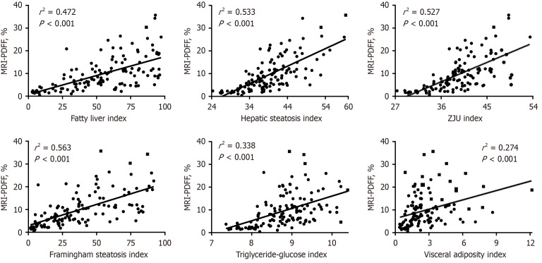
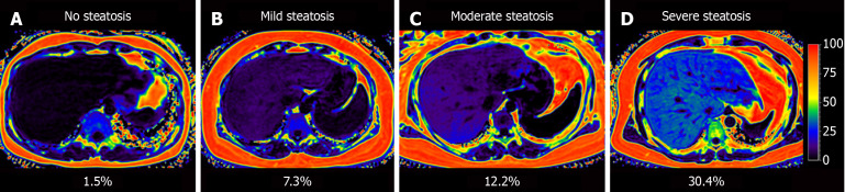
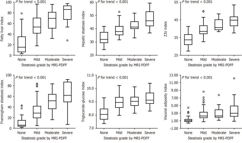
Methods: A total of 131 suspected NAFLD patients (60% male, median age 36 years) undergoing MRI-PDFF were consecutively recruited from a tertiary hospital. Steatosis grades determined by MRI-PDFF were classified as none (< 5%), mild (5%-11%), moderate (11%-17%), and severe (≥ 17%). Six steatosis biomarkers were calculated according to clinical parameters and laboratory tests, including fatty liver index, hepatic steatosis index, ZJU index, Framingham steatosis index, triglycerides and glucose index, and visceral adiposity index. The accuracy of these biomarkers in detecting hepatic steatosis was evaluated using the area under the receiver operating characteristic curves (AUCs). The Youden index was used to determine the optimal cut-off for each biomarker. The linear trend analysis of each biomarker across the steatosis grades was conducted by Mantel-Haenszel χ2 test. Spearman's rank correlation assessed the relationship between steatosis biomarkers and MRI-PDFF.
Results: Steatosis grades based on MRI-PDFF prevalence were: None 27%, mild 40%, moderate 15% and severe 18%. Six steatosis biomarkers showed a linear trend across the steatosis grades and a significant positive correlation with MRI-PDFF. The six steatosis biomarkers demonstrated AUCs near 0.90 (range: 0.857-0.912, all P < 0.001) for diagnosing NAFLD by MRI-PDFF ≥ 5%. The optimal cut-offs showed sensitivity between 84.4%-91.7% and specificity between 71.4%-85.7%. The diagnostic performance of these biomarkers in detecting moderate-to-severe and severe steatosis was relatively weaker.
Conclusion: These noninvasive steatosis biomarkers accurately diagnosed NAFLD and correlated well with MRI-PDFF for detecting NAFLD, but they did not effectively detect moderate or severe steatosis.

 求助内容:
求助内容: 应助结果提醒方式:


