{"title":"人工智能在基于图像的血肿增大预测中的表现:系统回顾和荟萃分析。","authors":"Wenjing Fan, Zhiping Wu, Wangyang Zhao, Luzhu Jia, Shuze Li, Wei Wei, Xin Chen","doi":"10.1080/07853890.2025.2515473","DOIUrl":null,"url":null,"abstract":"<p><strong>Background: </strong>Accurately predicting hematoma enlargement (HE) is crucial for improving the prognosis of patients with cerebral haemorrhage. Artificial intelligence (AI) is a potentially reliable assistant for medical image recognition. This study systematically reviews medical imaging articles on the predictive performance of AI in HE.</p><p><strong>Materials and methods: </strong>Retrieved relevant studies published before October, 2024 from Embase, Institute of Electrical and Electronics Engineers (IEEE), PubMed, Web of Science, and Cochrane Library databases. The diagnostic test of predicting hematoma enlargement based on CT image training artificial intelligence model, and reported 2 × 2 contingency tables or provided sensitivity (SE) and specificity (SP) for calculation. Two reviewers independently screened the retrieved citations and extracted data. The methodological quality of studies was assessed using the QUADAS-AI, and Preferred Reporting Items for Systematic reviews and Meta-Analyses was used to ensure standardised reporting of studies. Subgroup analysis was performed based on sample size, risk of bias, year of publication, ratio of training set to test set, and number of centres involved.</p><p><strong>Results: </strong>36 articles were included in this Systematic review to qualitative analysis, of which 23 have sufficient information for further quantitative analysis. Among these articles, there are a total of 7 articles used deep learning (DL) and 16 articles used machine learning (ML). The comprehensive SE and SP of ML are 78% (95% CI: 69-85%) and 85% (78-90%), respectively, while the AUC is 0.89 (0.86-0.91). The SE and SP of DL was 87% (95% CI: 80-92%) and 75% (67-81%), respectively, with an AUC of 0.88 (0.85-0.91). The subgroup analysis found that when the ratio of the training set to the test set is 7:3, the sensitivity is 0.77(0.62-0.91), <i>p</i> = 0.03; In terms of specificity, the group with sample size more than 200 has higher specificity, which is 0.83 (0.75-0.92), <i>p</i> = 0.02; among the risk groups in the study design, the specificity of the risk group was higher, which was 0.83 (0.76-0.89), <i>p</i> = 0.02. The group specificity of articles published before 2021 was higher, 0.84 (0.77-0.90); and the specificity of data from a single research centre was higher, which was 0.85 (0.80-0.91), <i>p</i> < 0.001.</p><p><strong>Conclusions: </strong>Artificial intelligence algorithms based on imaging have shown good performance in predicting HE.</p>","PeriodicalId":93874,"journal":{"name":"Annals of medicine","volume":"57 1","pages":"2515473"},"PeriodicalIF":4.3000,"publicationDate":"2025-12-01","publicationTypes":"Journal Article","fieldsOfStudy":null,"isOpenAccess":false,"openAccessPdf":"https://www.ncbi.nlm.nih.gov/pmc/articles/PMC12160331/pdf/","citationCount":"0","resultStr":"{\"title\":\"The performance of artificial intelligence in image-based prediction of hematoma enlargement: a systematic review and meta-analysis.\",\"authors\":\"Wenjing Fan, Zhiping Wu, Wangyang Zhao, Luzhu Jia, Shuze Li, Wei Wei, Xin Chen\",\"doi\":\"10.1080/07853890.2025.2515473\",\"DOIUrl\":null,\"url\":null,\"abstract\":\"<p><strong>Background: </strong>Accurately predicting hematoma enlargement (HE) is crucial for improving the prognosis of patients with cerebral haemorrhage. Artificial intelligence (AI) is a potentially reliable assistant for medical image recognition. This study systematically reviews medical imaging articles on the predictive performance of AI in HE.</p><p><strong>Materials and methods: </strong>Retrieved relevant studies published before October, 2024 from Embase, Institute of Electrical and Electronics Engineers (IEEE), PubMed, Web of Science, and Cochrane Library databases. The diagnostic test of predicting hematoma enlargement based on CT image training artificial intelligence model, and reported 2 × 2 contingency tables or provided sensitivity (SE) and specificity (SP) for calculation. Two reviewers independently screened the retrieved citations and extracted data. The methodological quality of studies was assessed using the QUADAS-AI, and Preferred Reporting Items for Systematic reviews and Meta-Analyses was used to ensure standardised reporting of studies. Subgroup analysis was performed based on sample size, risk of bias, year of publication, ratio of training set to test set, and number of centres involved.</p><p><strong>Results: </strong>36 articles were included in this Systematic review to qualitative analysis, of which 23 have sufficient information for further quantitative analysis. Among these articles, there are a total of 7 articles used deep learning (DL) and 16 articles used machine learning (ML). The comprehensive SE and SP of ML are 78% (95% CI: 69-85%) and 85% (78-90%), respectively, while the AUC is 0.89 (0.86-0.91). The SE and SP of DL was 87% (95% CI: 80-92%) and 75% (67-81%), respectively, with an AUC of 0.88 (0.85-0.91). The subgroup analysis found that when the ratio of the training set to the test set is 7:3, the sensitivity is 0.77(0.62-0.91), <i>p</i> = 0.03; In terms of specificity, the group with sample size more than 200 has higher specificity, which is 0.83 (0.75-0.92), <i>p</i> = 0.02; among the risk groups in the study design, the specificity of the risk group was higher, which was 0.83 (0.76-0.89), <i>p</i> = 0.02. The group specificity of articles published before 2021 was higher, 0.84 (0.77-0.90); and the specificity of data from a single research centre was higher, which was 0.85 (0.80-0.91), <i>p</i> < 0.001.</p><p><strong>Conclusions: </strong>Artificial intelligence algorithms based on imaging have shown good performance in predicting HE.</p>\",\"PeriodicalId\":93874,\"journal\":{\"name\":\"Annals of medicine\",\"volume\":\"57 1\",\"pages\":\"2515473\"},\"PeriodicalIF\":4.3000,\"publicationDate\":\"2025-12-01\",\"publicationTypes\":\"Journal Article\",\"fieldsOfStudy\":null,\"isOpenAccess\":false,\"openAccessPdf\":\"https://www.ncbi.nlm.nih.gov/pmc/articles/PMC12160331/pdf/\",\"citationCount\":\"0\",\"resultStr\":null,\"platform\":\"Semanticscholar\",\"paperid\":null,\"PeriodicalName\":\"Annals of medicine\",\"FirstCategoryId\":\"1085\",\"ListUrlMain\":\"https://doi.org/10.1080/07853890.2025.2515473\",\"RegionNum\":0,\"RegionCategory\":null,\"ArticlePicture\":[],\"TitleCN\":null,\"AbstractTextCN\":null,\"PMCID\":null,\"EPubDate\":\"2025/6/11 0:00:00\",\"PubModel\":\"Epub\",\"JCR\":\"\",\"JCRName\":\"\",\"Score\":null,\"Total\":0}","platform":"Semanticscholar","paperid":null,"PeriodicalName":"Annals of medicine","FirstCategoryId":"1085","ListUrlMain":"https://doi.org/10.1080/07853890.2025.2515473","RegionNum":0,"RegionCategory":null,"ArticlePicture":[],"TitleCN":null,"AbstractTextCN":null,"PMCID":null,"EPubDate":"2025/6/11 0:00:00","PubModel":"Epub","JCR":"","JCRName":"","Score":null,"Total":0}
The performance of artificial intelligence in image-based prediction of hematoma enlargement: a systematic review and meta-analysis.
Background: Accurately predicting hematoma enlargement (HE) is crucial for improving the prognosis of patients with cerebral haemorrhage. Artificial intelligence (AI) is a potentially reliable assistant for medical image recognition. This study systematically reviews medical imaging articles on the predictive performance of AI in HE.
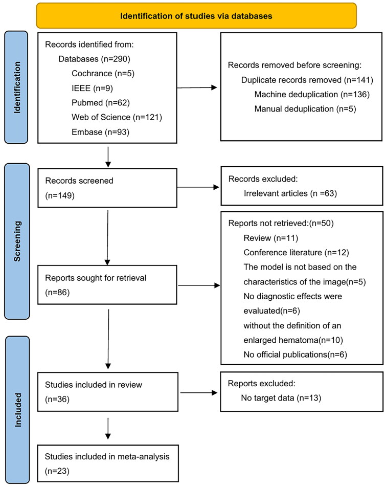
Materials and methods: Retrieved relevant studies published before October, 2024 from Embase, Institute of Electrical and Electronics Engineers (IEEE), PubMed, Web of Science, and Cochrane Library databases. The diagnostic test of predicting hematoma enlargement based on CT image training artificial intelligence model, and reported 2 × 2 contingency tables or provided sensitivity (SE) and specificity (SP) for calculation. Two reviewers independently screened the retrieved citations and extracted data. The methodological quality of studies was assessed using the QUADAS-AI, and Preferred Reporting Items for Systematic reviews and Meta-Analyses was used to ensure standardised reporting of studies. Subgroup analysis was performed based on sample size, risk of bias, year of publication, ratio of training set to test set, and number of centres involved.
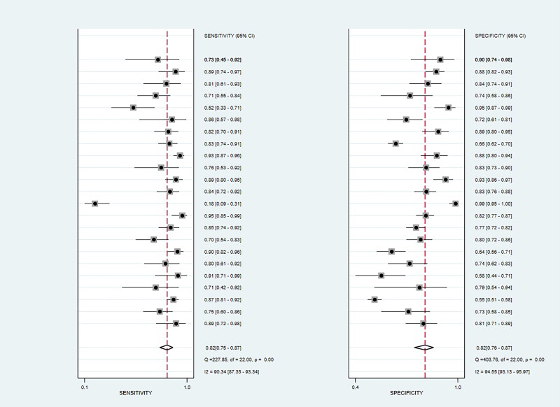
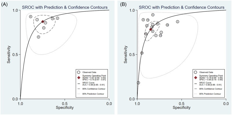
Results: 36 articles were included in this Systematic review to qualitative analysis, of which 23 have sufficient information for further quantitative analysis. Among these articles, there are a total of 7 articles used deep learning (DL) and 16 articles used machine learning (ML). The comprehensive SE and SP of ML are 78% (95% CI: 69-85%) and 85% (78-90%), respectively, while the AUC is 0.89 (0.86-0.91). The SE and SP of DL was 87% (95% CI: 80-92%) and 75% (67-81%), respectively, with an AUC of 0.88 (0.85-0.91). The subgroup analysis found that when the ratio of the training set to the test set is 7:3, the sensitivity is 0.77(0.62-0.91), p = 0.03; In terms of specificity, the group with sample size more than 200 has higher specificity, which is 0.83 (0.75-0.92), p = 0.02; among the risk groups in the study design, the specificity of the risk group was higher, which was 0.83 (0.76-0.89), p = 0.02. The group specificity of articles published before 2021 was higher, 0.84 (0.77-0.90); and the specificity of data from a single research centre was higher, which was 0.85 (0.80-0.91), p < 0.001.
Conclusions: Artificial intelligence algorithms based on imaging have shown good performance in predicting HE.

 求助内容:
求助内容: 应助结果提醒方式:


