Ammar K Alomran, Bandar A Alzahrani, Dana S Alamoud, Layan S Alsultan, Meshail M AlSaud, Raneem G Althobaiti, Badriah S Alruwaili
{"title":"术后x线在小儿桡骨远端骨折中的作用。","authors":"Ammar K Alomran, Bandar A Alzahrani, Dana S Alamoud, Layan S Alsultan, Meshail M AlSaud, Raneem G Althobaiti, Badriah S Alruwaili","doi":"10.5312/wjo.v16.i5.105590","DOIUrl":null,"url":null,"abstract":"<p><strong>Background: </strong>In pediatric age group patients (< 18 years old) treated operatively for distal radius/both bone fractures extending imaging beyond the initial postoperative period -particularly in uncomplicated cases - appears to provide limited additional benefit.</p><p><strong>Aim: </strong>To determine the necessary number of follow-up X-rays to use resources efficiently.</p><p><strong>Methods: </strong>Participants included in this study are pediatric age group patients who were treated operatively for distal radius/both bone fractures and were identified from a prospected collected data from the operating room database between the years 2009 and 2017. The data in the study included patients who had distal radius fractures and underwent fixation surgery (<i>n</i> = 88).</p><p><strong>Results: </strong>When assessing the difference in the odds of conducting 1 or less X-ray compared to 2 or more X-rays in regard to the type of fixation, the only significant difference is the closed reduction fixation method. Patients who underwent closed reduction method procedure have significantly lower odds of having 2 more X-rays compared to those who didn't have closed reduction method. Open reduction, internal fixation, and other fixation methods (close reduction and internal fixation, debridement, or epiphysiodesis) have higher odds of having two or more X-rays compared to patients who did not receive these methods; however, these odds are not statistically significant.</p><p><strong>Conclusion: </strong>The findings of this study reveal notable absence of a statistically significant association between the frequency of postoperative X-rays and the outcome of children with distal radius fractures.</p>","PeriodicalId":47843,"journal":{"name":"World Journal of Orthopedics","volume":"16 5","pages":"105590"},"PeriodicalIF":2.3000,"publicationDate":"2025-05-18","publicationTypes":"Journal Article","fieldsOfStudy":null,"isOpenAccess":false,"openAccessPdf":"https://www.ncbi.nlm.nih.gov/pmc/articles/PMC12146974/pdf/","citationCount":"0","resultStr":"{\"title\":\"Role of post-operative X-rays in distal-radius fractures among pediatric patients.\",\"authors\":\"Ammar K Alomran, Bandar A Alzahrani, Dana S Alamoud, Layan S Alsultan, Meshail M AlSaud, Raneem G Althobaiti, Badriah S Alruwaili\",\"doi\":\"10.5312/wjo.v16.i5.105590\",\"DOIUrl\":null,\"url\":null,\"abstract\":\"<p><strong>Background: </strong>In pediatric age group patients (< 18 years old) treated operatively for distal radius/both bone fractures extending imaging beyond the initial postoperative period -particularly in uncomplicated cases - appears to provide limited additional benefit.</p><p><strong>Aim: </strong>To determine the necessary number of follow-up X-rays to use resources efficiently.</p><p><strong>Methods: </strong>Participants included in this study are pediatric age group patients who were treated operatively for distal radius/both bone fractures and were identified from a prospected collected data from the operating room database between the years 2009 and 2017. The data in the study included patients who had distal radius fractures and underwent fixation surgery (<i>n</i> = 88).</p><p><strong>Results: </strong>When assessing the difference in the odds of conducting 1 or less X-ray compared to 2 or more X-rays in regard to the type of fixation, the only significant difference is the closed reduction fixation method. Patients who underwent closed reduction method procedure have significantly lower odds of having 2 more X-rays compared to those who didn't have closed reduction method. Open reduction, internal fixation, and other fixation methods (close reduction and internal fixation, debridement, or epiphysiodesis) have higher odds of having two or more X-rays compared to patients who did not receive these methods; however, these odds are not statistically significant.</p><p><strong>Conclusion: </strong>The findings of this study reveal notable absence of a statistically significant association between the frequency of postoperative X-rays and the outcome of children with distal radius fractures.</p>\",\"PeriodicalId\":47843,\"journal\":{\"name\":\"World Journal of Orthopedics\",\"volume\":\"16 5\",\"pages\":\"105590\"},\"PeriodicalIF\":2.3000,\"publicationDate\":\"2025-05-18\",\"publicationTypes\":\"Journal Article\",\"fieldsOfStudy\":null,\"isOpenAccess\":false,\"openAccessPdf\":\"https://www.ncbi.nlm.nih.gov/pmc/articles/PMC12146974/pdf/\",\"citationCount\":\"0\",\"resultStr\":null,\"platform\":\"Semanticscholar\",\"paperid\":null,\"PeriodicalName\":\"World Journal of Orthopedics\",\"FirstCategoryId\":\"1085\",\"ListUrlMain\":\"https://doi.org/10.5312/wjo.v16.i5.105590\",\"RegionNum\":0,\"RegionCategory\":null,\"ArticlePicture\":[],\"TitleCN\":null,\"AbstractTextCN\":null,\"PMCID\":null,\"EPubDate\":\"\",\"PubModel\":\"\",\"JCR\":\"Q2\",\"JCRName\":\"ORTHOPEDICS\",\"Score\":null,\"Total\":0}","platform":"Semanticscholar","paperid":null,"PeriodicalName":"World Journal of Orthopedics","FirstCategoryId":"1085","ListUrlMain":"https://doi.org/10.5312/wjo.v16.i5.105590","RegionNum":0,"RegionCategory":null,"ArticlePicture":[],"TitleCN":null,"AbstractTextCN":null,"PMCID":null,"EPubDate":"","PubModel":"","JCR":"Q2","JCRName":"ORTHOPEDICS","Score":null,"Total":0}
Role of post-operative X-rays in distal-radius fractures among pediatric patients.
Background: In pediatric age group patients (< 18 years old) treated operatively for distal radius/both bone fractures extending imaging beyond the initial postoperative period -particularly in uncomplicated cases - appears to provide limited additional benefit.
Aim: To determine the necessary number of follow-up X-rays to use resources efficiently.
Methods: Participants included in this study are pediatric age group patients who were treated operatively for distal radius/both bone fractures and were identified from a prospected collected data from the operating room database between the years 2009 and 2017. The data in the study included patients who had distal radius fractures and underwent fixation surgery (n = 88).
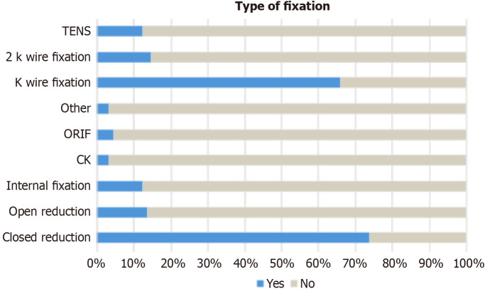
Results: When assessing the difference in the odds of conducting 1 or less X-ray compared to 2 or more X-rays in regard to the type of fixation, the only significant difference is the closed reduction fixation method. Patients who underwent closed reduction method procedure have significantly lower odds of having 2 more X-rays compared to those who didn't have closed reduction method. Open reduction, internal fixation, and other fixation methods (close reduction and internal fixation, debridement, or epiphysiodesis) have higher odds of having two or more X-rays compared to patients who did not receive these methods; however, these odds are not statistically significant.
Conclusion: The findings of this study reveal notable absence of a statistically significant association between the frequency of postoperative X-rays and the outcome of children with distal radius fractures.

 求助内容:
求助内容: 应助结果提醒方式:


