{"title":"肱骨髁上骨折患者肘关节x线测量与健康对照的比较分析。","authors":"Nuri K Ülgen, Batuhan Gencer, Özgür Doğan","doi":"10.5312/wjo.v16.i5.105734","DOIUrl":null,"url":null,"abstract":"<p><strong>Background: </strong>Supracondylar humeral fractures (SCHF) are the second most common fractures in childhood and can lead to short- and long-term complications. Despite their prevalence, the anatomical factors that predispose children to SCHF remain unclear. This study aimed to determine whether there are significant morphological differences in the elbow by comparing the radiographic angular measurements of the contralateral elbows of patients with SCHF to those of patients with distal radius fractures (DRF) and a healthy control group. We sought to explore if these morphological variations contribute to the occurrence of SCHF.</p><p><strong>Aim: </strong>To determine radiological parameters that may predispose to pediatric elbow fractures.</p><p><strong>Methods: </strong>Radiographs of 78 SCHF patients were analyzed for angular measurements of the contralateral elbow. Two control groups were used: 98 healthy children and 40 patients with DRF. Angular measurements included Baumann angle (BA), humeroulnar angle (HUA), humerus metaphysis-diaphysis angle (HMDA), humerus shaft-condylar angle (HSCA), and lateral capitellohumeral angle. Only BA, HUA, and HMDA were measured in the DRF group. Statistical analysis was performed to compare differences among groups.</p><p><strong>Results: </strong>Significant differences were found in elbow measurements between SCHF and control groups (<i>P</i> < 0.05). However, the mean values for all groups fell within the ranges described in the literature.</p><p><strong>Conclusion: </strong>While statistically significant differences were found in elbow morphology between SCHF patients and controls, these differences don't translate into clinically meaningful morphological deviations.</p>","PeriodicalId":47843,"journal":{"name":"World Journal of Orthopedics","volume":"16 5","pages":"105734"},"PeriodicalIF":2.3000,"publicationDate":"2025-05-18","publicationTypes":"Journal Article","fieldsOfStudy":null,"isOpenAccess":false,"openAccessPdf":"https://www.ncbi.nlm.nih.gov/pmc/articles/PMC12146972/pdf/","citationCount":"0","resultStr":"{\"title\":\"Comparative analysis of elbow radiographic measurements in patients with supracondylar humerus fractures and healthy controls.\",\"authors\":\"Nuri K Ülgen, Batuhan Gencer, Özgür Doğan\",\"doi\":\"10.5312/wjo.v16.i5.105734\",\"DOIUrl\":null,\"url\":null,\"abstract\":\"<p><strong>Background: </strong>Supracondylar humeral fractures (SCHF) are the second most common fractures in childhood and can lead to short- and long-term complications. Despite their prevalence, the anatomical factors that predispose children to SCHF remain unclear. This study aimed to determine whether there are significant morphological differences in the elbow by comparing the radiographic angular measurements of the contralateral elbows of patients with SCHF to those of patients with distal radius fractures (DRF) and a healthy control group. We sought to explore if these morphological variations contribute to the occurrence of SCHF.</p><p><strong>Aim: </strong>To determine radiological parameters that may predispose to pediatric elbow fractures.</p><p><strong>Methods: </strong>Radiographs of 78 SCHF patients were analyzed for angular measurements of the contralateral elbow. Two control groups were used: 98 healthy children and 40 patients with DRF. Angular measurements included Baumann angle (BA), humeroulnar angle (HUA), humerus metaphysis-diaphysis angle (HMDA), humerus shaft-condylar angle (HSCA), and lateral capitellohumeral angle. Only BA, HUA, and HMDA were measured in the DRF group. Statistical analysis was performed to compare differences among groups.</p><p><strong>Results: </strong>Significant differences were found in elbow measurements between SCHF and control groups (<i>P</i> < 0.05). However, the mean values for all groups fell within the ranges described in the literature.</p><p><strong>Conclusion: </strong>While statistically significant differences were found in elbow morphology between SCHF patients and controls, these differences don't translate into clinically meaningful morphological deviations.</p>\",\"PeriodicalId\":47843,\"journal\":{\"name\":\"World Journal of Orthopedics\",\"volume\":\"16 5\",\"pages\":\"105734\"},\"PeriodicalIF\":2.3000,\"publicationDate\":\"2025-05-18\",\"publicationTypes\":\"Journal Article\",\"fieldsOfStudy\":null,\"isOpenAccess\":false,\"openAccessPdf\":\"https://www.ncbi.nlm.nih.gov/pmc/articles/PMC12146972/pdf/\",\"citationCount\":\"0\",\"resultStr\":null,\"platform\":\"Semanticscholar\",\"paperid\":null,\"PeriodicalName\":\"World Journal of Orthopedics\",\"FirstCategoryId\":\"1085\",\"ListUrlMain\":\"https://doi.org/10.5312/wjo.v16.i5.105734\",\"RegionNum\":0,\"RegionCategory\":null,\"ArticlePicture\":[],\"TitleCN\":null,\"AbstractTextCN\":null,\"PMCID\":null,\"EPubDate\":\"\",\"PubModel\":\"\",\"JCR\":\"Q2\",\"JCRName\":\"ORTHOPEDICS\",\"Score\":null,\"Total\":0}","platform":"Semanticscholar","paperid":null,"PeriodicalName":"World Journal of Orthopedics","FirstCategoryId":"1085","ListUrlMain":"https://doi.org/10.5312/wjo.v16.i5.105734","RegionNum":0,"RegionCategory":null,"ArticlePicture":[],"TitleCN":null,"AbstractTextCN":null,"PMCID":null,"EPubDate":"","PubModel":"","JCR":"Q2","JCRName":"ORTHOPEDICS","Score":null,"Total":0}
Comparative analysis of elbow radiographic measurements in patients with supracondylar humerus fractures and healthy controls.
Background: Supracondylar humeral fractures (SCHF) are the second most common fractures in childhood and can lead to short- and long-term complications. Despite their prevalence, the anatomical factors that predispose children to SCHF remain unclear. This study aimed to determine whether there are significant morphological differences in the elbow by comparing the radiographic angular measurements of the contralateral elbows of patients with SCHF to those of patients with distal radius fractures (DRF) and a healthy control group. We sought to explore if these morphological variations contribute to the occurrence of SCHF.
Aim: To determine radiological parameters that may predispose to pediatric elbow fractures.
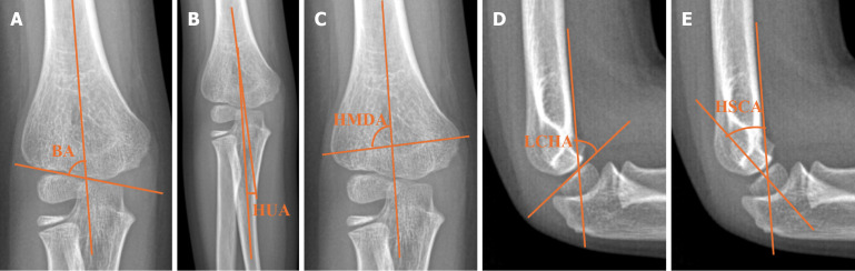
Methods: Radiographs of 78 SCHF patients were analyzed for angular measurements of the contralateral elbow. Two control groups were used: 98 healthy children and 40 patients with DRF. Angular measurements included Baumann angle (BA), humeroulnar angle (HUA), humerus metaphysis-diaphysis angle (HMDA), humerus shaft-condylar angle (HSCA), and lateral capitellohumeral angle. Only BA, HUA, and HMDA were measured in the DRF group. Statistical analysis was performed to compare differences among groups.
Results: Significant differences were found in elbow measurements between SCHF and control groups (P < 0.05). However, the mean values for all groups fell within the ranges described in the literature.
Conclusion: While statistically significant differences were found in elbow morphology between SCHF patients and controls, these differences don't translate into clinically meaningful morphological deviations.

 求助内容:
求助内容: 应助结果提醒方式:


