Michael Bambrick, Deena Godfrey, Mark D Johnson, Jeffrey D Esko, Biswa Choudhury, Alejandro Gomez Toledo, Mousumi Paulchakrabarti, Carla Fortes, Kevin J Staley, Ann-Christine Duhaime
{"title":"人创伤性脑损伤后细胞外基质成分的释放。","authors":"Michael Bambrick, Deena Godfrey, Mark D Johnson, Jeffrey D Esko, Biswa Choudhury, Alejandro Gomez Toledo, Mousumi Paulchakrabarti, Carla Fortes, Kevin J Staley, Ann-Christine Duhaime","doi":"10.1523/ENEURO.0488-24.2025","DOIUrl":null,"url":null,"abstract":"<p><p>Animal studies and human tissue experiments have demonstrated that traumatic brain injury (TBI) causes damage to the extracellular matrix (ECM). To test the hypothesis that TBI causes disruption of sulfated glycosaminoglycan (sGAG) in the ECM, we measured levels of sGAG in the cerebrospinal fluid (CSF), blood, and urine, in patients with severe TBI in the acute postinjury period. Samples of CSF, blood, and urine were obtained within 72 h of injury in patients who received external ventricular drains as part of their treatment of severe TBI. Levels of chondroitin and heparan sGAGs were measured, along with their disaccharide constituents. Demographic information, presence of polytrauma, brain injury load, and distance of radiologically visible parenchymal injury from the ventricle were analyzed for correlation with total subtype sGAG levels. Levels were measured in 14 patients ranging in age from 17 to 90 years. CSF sGAG levels were variable among patients, with higher sGAG levels in plasma compared with CSF. Patients with polytrauma had nonsignificantly higher blood sGAG compared with patients with isolated head injury. Subcategories of CSF sGAG levels correlated with distance from the ventricle of parenchymal injury but not with brain injury load. This study is the first to measure sGAG levels in ventricular CSF and the first to analyze levels in TBI. These data demonstrate the elevation locally of intracranial sGAGs after severe TBI and suggest rapid local metabolism of these breakdown products. The consequences of ECM breakdown may provide unique therapeutic and preventive avenues to mitigate postinjury sequelae.</p>","PeriodicalId":11617,"journal":{"name":"eNeuro","volume":" ","pages":""},"PeriodicalIF":2.7000,"publicationDate":"2025-06-26","publicationTypes":"Journal Article","fieldsOfStudy":null,"isOpenAccess":false,"openAccessPdf":"https://www.ncbi.nlm.nih.gov/pmc/articles/PMC12203761/pdf/","citationCount":"0","resultStr":"{\"title\":\"Release of Extracellular Matrix Components after Human Traumatic Brain Injury.\",\"authors\":\"Michael Bambrick, Deena Godfrey, Mark D Johnson, Jeffrey D Esko, Biswa Choudhury, Alejandro Gomez Toledo, Mousumi Paulchakrabarti, Carla Fortes, Kevin J Staley, Ann-Christine Duhaime\",\"doi\":\"10.1523/ENEURO.0488-24.2025\",\"DOIUrl\":null,\"url\":null,\"abstract\":\"<p><p>Animal studies and human tissue experiments have demonstrated that traumatic brain injury (TBI) causes damage to the extracellular matrix (ECM). To test the hypothesis that TBI causes disruption of sulfated glycosaminoglycan (sGAG) in the ECM, we measured levels of sGAG in the cerebrospinal fluid (CSF), blood, and urine, in patients with severe TBI in the acute postinjury period. Samples of CSF, blood, and urine were obtained within 72 h of injury in patients who received external ventricular drains as part of their treatment of severe TBI. Levels of chondroitin and heparan sGAGs were measured, along with their disaccharide constituents. Demographic information, presence of polytrauma, brain injury load, and distance of radiologically visible parenchymal injury from the ventricle were analyzed for correlation with total subtype sGAG levels. Levels were measured in 14 patients ranging in age from 17 to 90 years. CSF sGAG levels were variable among patients, with higher sGAG levels in plasma compared with CSF. Patients with polytrauma had nonsignificantly higher blood sGAG compared with patients with isolated head injury. Subcategories of CSF sGAG levels correlated with distance from the ventricle of parenchymal injury but not with brain injury load. This study is the first to measure sGAG levels in ventricular CSF and the first to analyze levels in TBI. These data demonstrate the elevation locally of intracranial sGAGs after severe TBI and suggest rapid local metabolism of these breakdown products. The consequences of ECM breakdown may provide unique therapeutic and preventive avenues to mitigate postinjury sequelae.</p>\",\"PeriodicalId\":11617,\"journal\":{\"name\":\"eNeuro\",\"volume\":\" \",\"pages\":\"\"},\"PeriodicalIF\":2.7000,\"publicationDate\":\"2025-06-26\",\"publicationTypes\":\"Journal Article\",\"fieldsOfStudy\":null,\"isOpenAccess\":false,\"openAccessPdf\":\"https://www.ncbi.nlm.nih.gov/pmc/articles/PMC12203761/pdf/\",\"citationCount\":\"0\",\"resultStr\":null,\"platform\":\"Semanticscholar\",\"paperid\":null,\"PeriodicalName\":\"eNeuro\",\"FirstCategoryId\":\"3\",\"ListUrlMain\":\"https://doi.org/10.1523/ENEURO.0488-24.2025\",\"RegionNum\":3,\"RegionCategory\":\"医学\",\"ArticlePicture\":[],\"TitleCN\":null,\"AbstractTextCN\":null,\"PMCID\":null,\"EPubDate\":\"2025/6/1 0:00:00\",\"PubModel\":\"Print\",\"JCR\":\"Q3\",\"JCRName\":\"NEUROSCIENCES\",\"Score\":null,\"Total\":0}","platform":"Semanticscholar","paperid":null,"PeriodicalName":"eNeuro","FirstCategoryId":"3","ListUrlMain":"https://doi.org/10.1523/ENEURO.0488-24.2025","RegionNum":3,"RegionCategory":"医学","ArticlePicture":[],"TitleCN":null,"AbstractTextCN":null,"PMCID":null,"EPubDate":"2025/6/1 0:00:00","PubModel":"Print","JCR":"Q3","JCRName":"NEUROSCIENCES","Score":null,"Total":0}
Release of Extracellular Matrix Components after Human Traumatic Brain Injury.
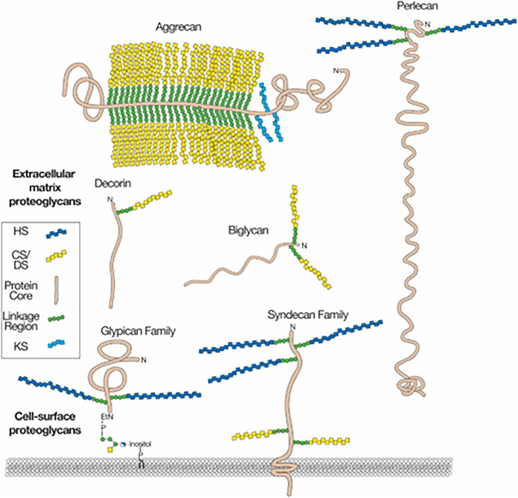
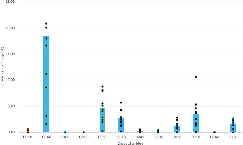
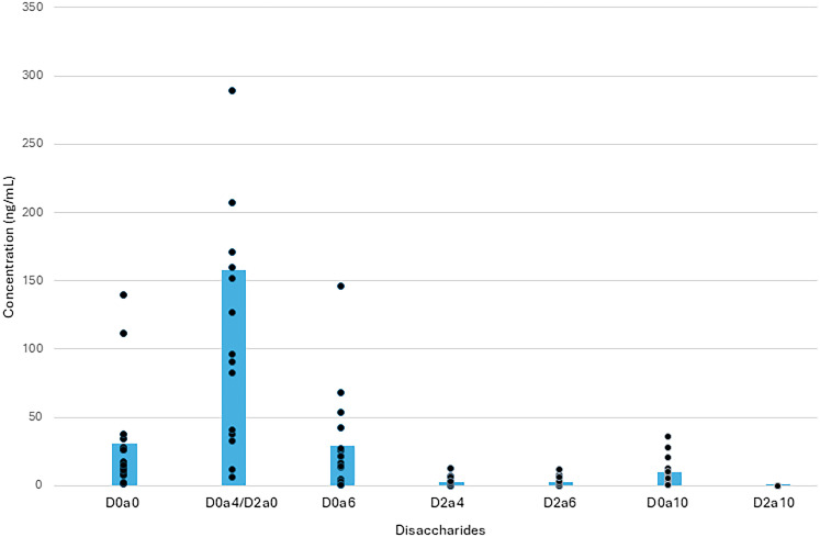
Animal studies and human tissue experiments have demonstrated that traumatic brain injury (TBI) causes damage to the extracellular matrix (ECM). To test the hypothesis that TBI causes disruption of sulfated glycosaminoglycan (sGAG) in the ECM, we measured levels of sGAG in the cerebrospinal fluid (CSF), blood, and urine, in patients with severe TBI in the acute postinjury period. Samples of CSF, blood, and urine were obtained within 72 h of injury in patients who received external ventricular drains as part of their treatment of severe TBI. Levels of chondroitin and heparan sGAGs were measured, along with their disaccharide constituents. Demographic information, presence of polytrauma, brain injury load, and distance of radiologically visible parenchymal injury from the ventricle were analyzed for correlation with total subtype sGAG levels. Levels were measured in 14 patients ranging in age from 17 to 90 years. CSF sGAG levels were variable among patients, with higher sGAG levels in plasma compared with CSF. Patients with polytrauma had nonsignificantly higher blood sGAG compared with patients with isolated head injury. Subcategories of CSF sGAG levels correlated with distance from the ventricle of parenchymal injury but not with brain injury load. This study is the first to measure sGAG levels in ventricular CSF and the first to analyze levels in TBI. These data demonstrate the elevation locally of intracranial sGAGs after severe TBI and suggest rapid local metabolism of these breakdown products. The consequences of ECM breakdown may provide unique therapeutic and preventive avenues to mitigate postinjury sequelae.
期刊介绍:
An open-access journal from the Society for Neuroscience, eNeuro publishes high-quality, broad-based, peer-reviewed research focused solely on the field of neuroscience. eNeuro embodies an emerging scientific vision that offers a new experience for authors and readers, all in support of the Society’s mission to advance understanding of the brain and nervous system.
 求助内容:
求助内容: 应助结果提醒方式:


