{"title":"轻度至中度缺血性中风后的持续性视力损害。","authors":"Chamini Niroshika Wijesundera, Sheila Gillard Crewther, Tissa Wijeratne, Algis J Vingrys","doi":"10.3389/fopht.2025.1505836","DOIUrl":null,"url":null,"abstract":"<p><strong>Background: </strong>Vision is rarely appraised either acutely or during recovery, following acute ischemic stroke. Our previous study found significant deficits in visual function after 2 to 3 days in ~68% of hospitalized mild-to-moderate acute ischemic stroke (AIS) patients with no comorbid eye disease. The purpose of this study was to evaluate recovery in vision after 2-6 months in a subgroup of the original participants.</p><p><strong>Methods: </strong>Visual assessments were performed within the first week of admission and 2-6 months later. Testing was achieved on an iPad and included visual acuity (VA), VA-in-noise, visual field, visual neglect, and time to complete an eye-hand coordination (EHC) task. All cases were radiologically confirmed, and 10 had left hemisphere lesions. The outcomes were compared to 20 age-matched healthy controls who were tested and retested over a similar duration using the same vision tests. The testing took 12 min.</p><p><strong>Results: </strong>During the first week of admission, 19/20 (95%) AIS patients returned normal visual acuity (>6/12 VA, <i>p</i> = 0.11), yet 11/20 (55%) had reduced VA-in-noise (<i>p</i> < 0.000).Visual neglect was present in 2/20 cases. Visual field defects were present in 16/20 (80%, <i>p</i> < 0.001), with 7/16 (44%) being unaware of their visual field loss. All of the patients chose to use their dominant right hand despite 10 having left hemisphere lesions and 13/20 (65%, <i>p</i> < 0.001) returning longer times to complete the EHC tracing tasks. After 2-6 months of recovery, all stroke patients returned normal visual attention, although 3/20 (15%) continued to show reduced VA in the presence of noise masks. Seven out of 20 (35%) retained visual field defects, and 8/20 (40%, three right and five left hemisphere lesions) had visuomotor impairment. Posterior circulation territory strokes and left hemisphere lesions were more likely to result in a persistent visual field loss and visuomotor deficit.</p><p><strong>Conclusion: </strong>Given that stroke is the leading cause of neurological disability affecting over 110 million people, our findings highlight the necessity for both acute and longitudinal vision assessments subsequent to mild stroke. Exposing the persistent limitations in visual functions could aid in identifying suitability for driving and the visuomotor rehabilitation of stroke survivors.</p><p><strong>Clinical trial registration: </strong>https://www.ANZCTR.org.au/ACTRN12618001111268.aspx, identifier ACTRN12618001111268.</p>","PeriodicalId":73096,"journal":{"name":"Frontiers in ophthalmology","volume":"5 ","pages":"1505836"},"PeriodicalIF":0.9000,"publicationDate":"2025-05-26","publicationTypes":"Journal Article","fieldsOfStudy":null,"isOpenAccess":false,"openAccessPdf":"https://www.ncbi.nlm.nih.gov/pmc/articles/PMC12146193/pdf/","citationCount":"0","resultStr":"{\"title\":\"Persistent visual impairments following mild-to-moderate ischemic stroke.\",\"authors\":\"Chamini Niroshika Wijesundera, Sheila Gillard Crewther, Tissa Wijeratne, Algis J Vingrys\",\"doi\":\"10.3389/fopht.2025.1505836\",\"DOIUrl\":null,\"url\":null,\"abstract\":\"<p><strong>Background: </strong>Vision is rarely appraised either acutely or during recovery, following acute ischemic stroke. Our previous study found significant deficits in visual function after 2 to 3 days in ~68% of hospitalized mild-to-moderate acute ischemic stroke (AIS) patients with no comorbid eye disease. The purpose of this study was to evaluate recovery in vision after 2-6 months in a subgroup of the original participants.</p><p><strong>Methods: </strong>Visual assessments were performed within the first week of admission and 2-6 months later. Testing was achieved on an iPad and included visual acuity (VA), VA-in-noise, visual field, visual neglect, and time to complete an eye-hand coordination (EHC) task. All cases were radiologically confirmed, and 10 had left hemisphere lesions. The outcomes were compared to 20 age-matched healthy controls who were tested and retested over a similar duration using the same vision tests. The testing took 12 min.</p><p><strong>Results: </strong>During the first week of admission, 19/20 (95%) AIS patients returned normal visual acuity (>6/12 VA, <i>p</i> = 0.11), yet 11/20 (55%) had reduced VA-in-noise (<i>p</i> < 0.000).Visual neglect was present in 2/20 cases. Visual field defects were present in 16/20 (80%, <i>p</i> < 0.001), with 7/16 (44%) being unaware of their visual field loss. All of the patients chose to use their dominant right hand despite 10 having left hemisphere lesions and 13/20 (65%, <i>p</i> < 0.001) returning longer times to complete the EHC tracing tasks. After 2-6 months of recovery, all stroke patients returned normal visual attention, although 3/20 (15%) continued to show reduced VA in the presence of noise masks. Seven out of 20 (35%) retained visual field defects, and 8/20 (40%, three right and five left hemisphere lesions) had visuomotor impairment. Posterior circulation territory strokes and left hemisphere lesions were more likely to result in a persistent visual field loss and visuomotor deficit.</p><p><strong>Conclusion: </strong>Given that stroke is the leading cause of neurological disability affecting over 110 million people, our findings highlight the necessity for both acute and longitudinal vision assessments subsequent to mild stroke. Exposing the persistent limitations in visual functions could aid in identifying suitability for driving and the visuomotor rehabilitation of stroke survivors.</p><p><strong>Clinical trial registration: </strong>https://www.ANZCTR.org.au/ACTRN12618001111268.aspx, identifier ACTRN12618001111268.</p>\",\"PeriodicalId\":73096,\"journal\":{\"name\":\"Frontiers in ophthalmology\",\"volume\":\"5 \",\"pages\":\"1505836\"},\"PeriodicalIF\":0.9000,\"publicationDate\":\"2025-05-26\",\"publicationTypes\":\"Journal Article\",\"fieldsOfStudy\":null,\"isOpenAccess\":false,\"openAccessPdf\":\"https://www.ncbi.nlm.nih.gov/pmc/articles/PMC12146193/pdf/\",\"citationCount\":\"0\",\"resultStr\":null,\"platform\":\"Semanticscholar\",\"paperid\":null,\"PeriodicalName\":\"Frontiers in ophthalmology\",\"FirstCategoryId\":\"1085\",\"ListUrlMain\":\"https://doi.org/10.3389/fopht.2025.1505836\",\"RegionNum\":0,\"RegionCategory\":null,\"ArticlePicture\":[],\"TitleCN\":null,\"AbstractTextCN\":null,\"PMCID\":null,\"EPubDate\":\"2025/1/1 0:00:00\",\"PubModel\":\"eCollection\",\"JCR\":\"\",\"JCRName\":\"\",\"Score\":null,\"Total\":0}","platform":"Semanticscholar","paperid":null,"PeriodicalName":"Frontiers in ophthalmology","FirstCategoryId":"1085","ListUrlMain":"https://doi.org/10.3389/fopht.2025.1505836","RegionNum":0,"RegionCategory":null,"ArticlePicture":[],"TitleCN":null,"AbstractTextCN":null,"PMCID":null,"EPubDate":"2025/1/1 0:00:00","PubModel":"eCollection","JCR":"","JCRName":"","Score":null,"Total":0}
引用次数: 0
摘要
背景:在急性缺血性脑卒中后的急性期或恢复期,视力很少被评估。我们之前的研究发现,68%的住院的轻中度急性缺血性卒中(AIS)患者在2 - 3天后出现明显的视觉功能缺陷,且无眼部合并症。本研究的目的是评估原始参与者的亚组在2-6个月后的视力恢复情况。方法:在入院第一周及2-6个月后进行视力评估。测试在iPad上完成,包括视力(VA)、VA-in-noise、视野、视觉忽视和完成手眼协调(EHC)任务的时间。所有病例均经影像学证实,其中10例有左半球病变。结果与20名年龄匹配的健康对照者进行了比较,这些对照者在相似的时间内使用相同的视力测试进行了测试和重新测试。结果:入院第一周,19/20 (95%)AIS患者恢复正常视力(>6/ 12va, p = 0.11), 11/20(55%)患者噪声VA降低(p < 0.000)。2/20的病例存在视觉忽视。16/20 (80%, p < 0.001)存在视野缺损,7/16(44%)不知道自己的视野丧失。尽管10名患者有左半球病变,13/20 (65%,p < 0.001)返回更长时间来完成EHC追踪任务,但所有患者都选择使用他们的右手。恢复2-6个月后,所有脑卒中患者恢复了正常的视觉注意,尽管3/20(15%)的患者在噪音面罩的存在下继续表现出VA降低。20例患者中有7例(35%)存在视野缺损,8/20例(40%,3例右半球病变,5例左半球病变)存在视觉运动障碍。后循环领域中风和左半球病变更可能导致持续的视野丧失和视觉运动障碍。结论:鉴于中风是影响超过1.1亿人的神经功能障碍的主要原因,我们的研究结果强调了轻度中风后急性和纵向视力评估的必要性。揭示视觉功能的持续限制可以帮助中风幸存者识别驾驶和视觉运动康复的适宜性。临床试验注册:https://www.ANZCTR.org.au/ACTRN12618001111268.aspx,标识符ACTRN12618001111268。
Persistent visual impairments following mild-to-moderate ischemic stroke.
Background: Vision is rarely appraised either acutely or during recovery, following acute ischemic stroke. Our previous study found significant deficits in visual function after 2 to 3 days in ~68% of hospitalized mild-to-moderate acute ischemic stroke (AIS) patients with no comorbid eye disease. The purpose of this study was to evaluate recovery in vision after 2-6 months in a subgroup of the original participants.
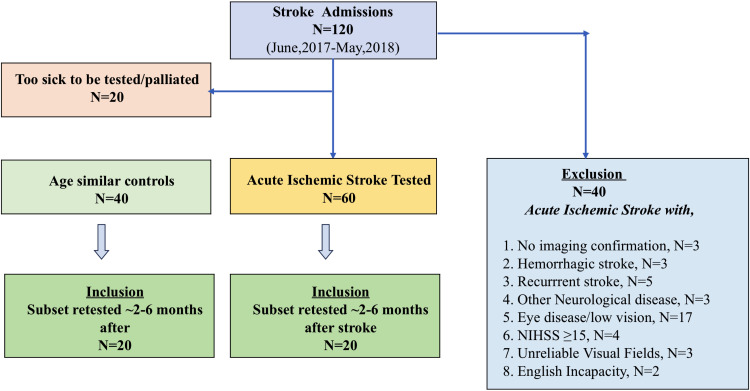
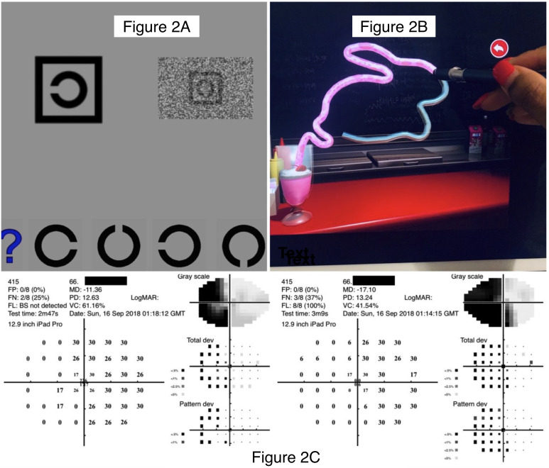
Methods: Visual assessments were performed within the first week of admission and 2-6 months later. Testing was achieved on an iPad and included visual acuity (VA), VA-in-noise, visual field, visual neglect, and time to complete an eye-hand coordination (EHC) task. All cases were radiologically confirmed, and 10 had left hemisphere lesions. The outcomes were compared to 20 age-matched healthy controls who were tested and retested over a similar duration using the same vision tests. The testing took 12 min.
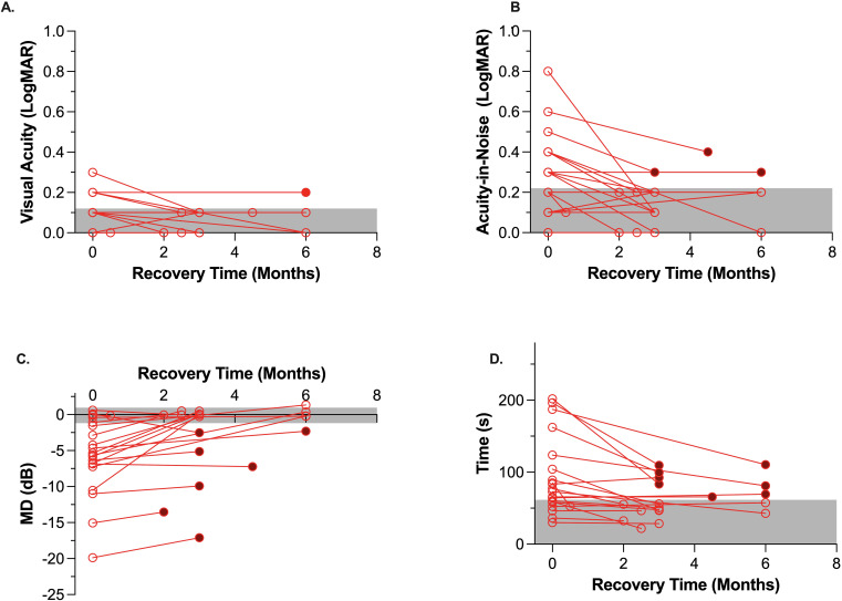
Results: During the first week of admission, 19/20 (95%) AIS patients returned normal visual acuity (>6/12 VA, p = 0.11), yet 11/20 (55%) had reduced VA-in-noise (p < 0.000).Visual neglect was present in 2/20 cases. Visual field defects were present in 16/20 (80%, p < 0.001), with 7/16 (44%) being unaware of their visual field loss. All of the patients chose to use their dominant right hand despite 10 having left hemisphere lesions and 13/20 (65%, p < 0.001) returning longer times to complete the EHC tracing tasks. After 2-6 months of recovery, all stroke patients returned normal visual attention, although 3/20 (15%) continued to show reduced VA in the presence of noise masks. Seven out of 20 (35%) retained visual field defects, and 8/20 (40%, three right and five left hemisphere lesions) had visuomotor impairment. Posterior circulation territory strokes and left hemisphere lesions were more likely to result in a persistent visual field loss and visuomotor deficit.
Conclusion: Given that stroke is the leading cause of neurological disability affecting over 110 million people, our findings highlight the necessity for both acute and longitudinal vision assessments subsequent to mild stroke. Exposing the persistent limitations in visual functions could aid in identifying suitability for driving and the visuomotor rehabilitation of stroke survivors.

 求助内容:
求助内容: 应助结果提醒方式:


