{"title":"在轻度创伤性脑损伤的年轻猪模型中,体积和弥散张量成像异常与脑震荡后的行为改变有关。","authors":"Islam Sanjida, Netzley Alesa, Li Chenyang, Zhang Jiangyang, Dávila-Montero Bianca, Vazquez Ana, Subbaiah Shaun, Meoded Avner, Munoz Kirk, Colbath Aimee, Huang Jie, Mejia-Alvarez Ricardo, Manfredi Jane, Pelled Galit","doi":"10.1002/nbm.70074","DOIUrl":null,"url":null,"abstract":"<p><p>Mild traumatic brain injury (mTBI) caused by sports-related incidents in children and youth often leads to prolonged cognitive impairments but remains difficult to diagnose. In order to identify clinically relevant imaging and behavioral biomarkers associated concussion, a closed-head mTBI was induced in adolescent pigs. Twelve (n = 4 male and n = 8 female), 16-week old Yucatan pigs were tested; n = 6 received mTBI and n = 6 received a sham procedure. T1-weighted imaging was used to assess volumetric alterations in different regions of the brain and diffusion tensor imaging (DTI) to examine microstructural damage in white matter. The pigs were imaged at 1- and 3-month post-injury. Neuropsychological screening for executive function and anxiety were performed before and in the months after the injury. The volumetric analysis showed significant longitudinal changes in pigs with mTBI compared with sham, which may be attributed to swelling and neuroinflammation. Fractional anisotropy (FA) values derived from DTI images demonstrated a 21% increase in corpus callosum from 1 to 3 months in mTBI pigs, which is significantly higher than in sham pigs (4.8%). Additionally, comparisons of the left and right internal capsules revealed a decrease in FA in the right internal capsule for mTBI pigs, which may indicate demyelination. The neuroimaging results suggest that the injury had disrupted the maturation of white and gray matter in the developing brain. Behavioral testing showed that compare to sham pigs, mTBI pigs exhibited 23% increased activity in open field tests, 35% incraesed escape attempts, along with a 65% decrease in interaction with the novel object, suggesting possible memory impairments and cognitive deficits. The correlation analysis showed an associations between volumetric features and behavioral metrics. Furthermore, a machine learning model, which integrated FA, volumetric features and behavioral test metrics, achieved 67% accuracy, indicating its potential to differentiate the two groups. Thus, the imaging biomarkers were indicative of long-term behavioral impairments and could be crucial to the clinical management of concussion in youth.</p>","PeriodicalId":19309,"journal":{"name":"NMR in Biomedicine","volume":"38 7","pages":"e70074"},"PeriodicalIF":2.7000,"publicationDate":"2025-07-01","publicationTypes":"Journal Article","fieldsOfStudy":null,"isOpenAccess":false,"openAccessPdf":"https://www.ncbi.nlm.nih.gov/pmc/articles/PMC12149694/pdf/","citationCount":"0","resultStr":"{\"title\":\"Volumetric and Diffusion Tensor Imaging Abnormalities Are Associated With Behavioral Changes Post-Concussion in a Youth Pig Model of Mild Traumatic Brain Injury.\",\"authors\":\"Islam Sanjida, Netzley Alesa, Li Chenyang, Zhang Jiangyang, Dávila-Montero Bianca, Vazquez Ana, Subbaiah Shaun, Meoded Avner, Munoz Kirk, Colbath Aimee, Huang Jie, Mejia-Alvarez Ricardo, Manfredi Jane, Pelled Galit\",\"doi\":\"10.1002/nbm.70074\",\"DOIUrl\":null,\"url\":null,\"abstract\":\"<p><p>Mild traumatic brain injury (mTBI) caused by sports-related incidents in children and youth often leads to prolonged cognitive impairments but remains difficult to diagnose. In order to identify clinically relevant imaging and behavioral biomarkers associated concussion, a closed-head mTBI was induced in adolescent pigs. Twelve (n = 4 male and n = 8 female), 16-week old Yucatan pigs were tested; n = 6 received mTBI and n = 6 received a sham procedure. T1-weighted imaging was used to assess volumetric alterations in different regions of the brain and diffusion tensor imaging (DTI) to examine microstructural damage in white matter. The pigs were imaged at 1- and 3-month post-injury. Neuropsychological screening for executive function and anxiety were performed before and in the months after the injury. The volumetric analysis showed significant longitudinal changes in pigs with mTBI compared with sham, which may be attributed to swelling and neuroinflammation. Fractional anisotropy (FA) values derived from DTI images demonstrated a 21% increase in corpus callosum from 1 to 3 months in mTBI pigs, which is significantly higher than in sham pigs (4.8%). Additionally, comparisons of the left and right internal capsules revealed a decrease in FA in the right internal capsule for mTBI pigs, which may indicate demyelination. The neuroimaging results suggest that the injury had disrupted the maturation of white and gray matter in the developing brain. Behavioral testing showed that compare to sham pigs, mTBI pigs exhibited 23% increased activity in open field tests, 35% incraesed escape attempts, along with a 65% decrease in interaction with the novel object, suggesting possible memory impairments and cognitive deficits. The correlation analysis showed an associations between volumetric features and behavioral metrics. Furthermore, a machine learning model, which integrated FA, volumetric features and behavioral test metrics, achieved 67% accuracy, indicating its potential to differentiate the two groups. Thus, the imaging biomarkers were indicative of long-term behavioral impairments and could be crucial to the clinical management of concussion in youth.</p>\",\"PeriodicalId\":19309,\"journal\":{\"name\":\"NMR in Biomedicine\",\"volume\":\"38 7\",\"pages\":\"e70074\"},\"PeriodicalIF\":2.7000,\"publicationDate\":\"2025-07-01\",\"publicationTypes\":\"Journal Article\",\"fieldsOfStudy\":null,\"isOpenAccess\":false,\"openAccessPdf\":\"https://www.ncbi.nlm.nih.gov/pmc/articles/PMC12149694/pdf/\",\"citationCount\":\"0\",\"resultStr\":null,\"platform\":\"Semanticscholar\",\"paperid\":null,\"PeriodicalName\":\"NMR in Biomedicine\",\"FirstCategoryId\":\"3\",\"ListUrlMain\":\"https://doi.org/10.1002/nbm.70074\",\"RegionNum\":4,\"RegionCategory\":\"医学\",\"ArticlePicture\":[],\"TitleCN\":null,\"AbstractTextCN\":null,\"PMCID\":null,\"EPubDate\":\"\",\"PubModel\":\"\",\"JCR\":\"Q2\",\"JCRName\":\"BIOPHYSICS\",\"Score\":null,\"Total\":0}","platform":"Semanticscholar","paperid":null,"PeriodicalName":"NMR in Biomedicine","FirstCategoryId":"3","ListUrlMain":"https://doi.org/10.1002/nbm.70074","RegionNum":4,"RegionCategory":"医学","ArticlePicture":[],"TitleCN":null,"AbstractTextCN":null,"PMCID":null,"EPubDate":"","PubModel":"","JCR":"Q2","JCRName":"BIOPHYSICS","Score":null,"Total":0}
Volumetric and Diffusion Tensor Imaging Abnormalities Are Associated With Behavioral Changes Post-Concussion in a Youth Pig Model of Mild Traumatic Brain Injury.
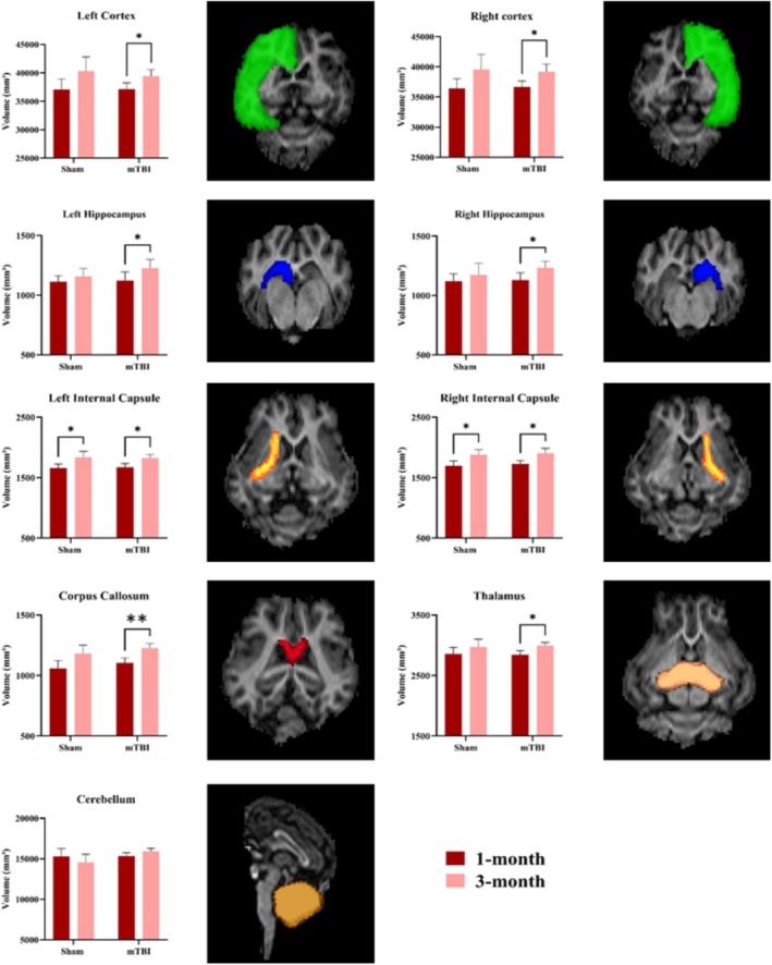
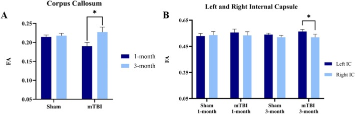
Mild traumatic brain injury (mTBI) caused by sports-related incidents in children and youth often leads to prolonged cognitive impairments but remains difficult to diagnose. In order to identify clinically relevant imaging and behavioral biomarkers associated concussion, a closed-head mTBI was induced in adolescent pigs. Twelve (n = 4 male and n = 8 female), 16-week old Yucatan pigs were tested; n = 6 received mTBI and n = 6 received a sham procedure. T1-weighted imaging was used to assess volumetric alterations in different regions of the brain and diffusion tensor imaging (DTI) to examine microstructural damage in white matter. The pigs were imaged at 1- and 3-month post-injury. Neuropsychological screening for executive function and anxiety were performed before and in the months after the injury. The volumetric analysis showed significant longitudinal changes in pigs with mTBI compared with sham, which may be attributed to swelling and neuroinflammation. Fractional anisotropy (FA) values derived from DTI images demonstrated a 21% increase in corpus callosum from 1 to 3 months in mTBI pigs, which is significantly higher than in sham pigs (4.8%). Additionally, comparisons of the left and right internal capsules revealed a decrease in FA in the right internal capsule for mTBI pigs, which may indicate demyelination. The neuroimaging results suggest that the injury had disrupted the maturation of white and gray matter in the developing brain. Behavioral testing showed that compare to sham pigs, mTBI pigs exhibited 23% increased activity in open field tests, 35% incraesed escape attempts, along with a 65% decrease in interaction with the novel object, suggesting possible memory impairments and cognitive deficits. The correlation analysis showed an associations between volumetric features and behavioral metrics. Furthermore, a machine learning model, which integrated FA, volumetric features and behavioral test metrics, achieved 67% accuracy, indicating its potential to differentiate the two groups. Thus, the imaging biomarkers were indicative of long-term behavioral impairments and could be crucial to the clinical management of concussion in youth.
期刊介绍:
NMR in Biomedicine is a journal devoted to the publication of original full-length papers, rapid communications and review articles describing the development of magnetic resonance spectroscopy or imaging methods or their use to investigate physiological, biochemical, biophysical or medical problems. Topics for submitted papers should be in one of the following general categories: (a) development of methods and instrumentation for MR of biological systems; (b) studies of normal or diseased organs, tissues or cells; (c) diagnosis or treatment of disease. Reports may cover work on patients or healthy human subjects, in vivo animal experiments, studies of isolated organs or cultured cells, analysis of tissue extracts, NMR theory, experimental techniques, or instrumentation.

 求助内容:
求助内容: 应助结果提醒方式:


