Emel Olga Onay, Derin Bugu Yuzer, Eda Cakmak, Kamran Gulsahi
{"title":"锥束计算机断层扫描和根管医师水平对根吸收诊断和治疗计划的影响。","authors":"Emel Olga Onay, Derin Bugu Yuzer, Eda Cakmak, Kamran Gulsahi","doi":"10.4317/jced.62382","DOIUrl":null,"url":null,"abstract":"<p><strong>Background: </strong>A precise diagnosis is essential for formulating an effective treatment plan for both internal and external resorptions. Cone-beam computed tomography (CBCT) offers a three-dimensional view of the maxillofacial area, capturing images from coronal, axial, and sagittal angles. This method overcomes the limitations of conventional intraoral radiography (IR), especially when it comes to detecting and identifying defects related to internal and external resorption. Thus, the aim of this study was to assess whether CBCT imaging affects the accuracy of diagnosing and planning treatment for internal and external resorption defects differently between endodontic residents (ERs) and specialists (ESs).</p><p><strong>Material and methods: </strong>Thirty-five clinicians reviewed 3 internal and 3 external resorption cases using clinical histories and intraoral radiographs (IRs), then answered questions about their diagnosis and treatment decisions. One month later, they re-evaluated the cases with CBCT and answered similar questions. Data analyzed using Mc-Nemar chi-square test and Prevalence Adjusted Bias Adjusted Kappa statistic. The level of statistical significance was set off <i>p</i>< 0.05 in all data.</p><p><strong>Results: </strong>CBCT significantly improved diagnostic accuracy in 2 out of 6 cases (<i>p</i>< 0.001) and altered the treatment plan in 4 out of 6 cases (<i>p</i>< 0.05). There was no significant difference between ERs and ESs regarding diagnosis and treatment planning using the same imaging technique (<i>p</i>> 0.05).</p><p><strong>Conclusions: </strong>This study suggests that CBCT provides more detailed information compared to IR, with both imaging techniques allowing ERs and ESs to achieve similar diagnostic and treatment planning accuracy. <b>Key words:</b>Clinical decision making, cone-beam computed tomography, dental radiography, diagnosis, root resorption.</p>","PeriodicalId":15376,"journal":{"name":"Journal of Clinical and Experimental Dentistry","volume":"17 5","pages":"e515-e520"},"PeriodicalIF":0.0000,"publicationDate":"2025-05-01","publicationTypes":"Journal Article","fieldsOfStudy":null,"isOpenAccess":false,"openAccessPdf":"https://www.ncbi.nlm.nih.gov/pmc/articles/PMC12142370/pdf/","citationCount":"0","resultStr":"{\"title\":\"The influence of cone-beam computed tomography and endodontic practitioners' proficiency level on diagnosis and treatment planning of root resorption.\",\"authors\":\"Emel Olga Onay, Derin Bugu Yuzer, Eda Cakmak, Kamran Gulsahi\",\"doi\":\"10.4317/jced.62382\",\"DOIUrl\":null,\"url\":null,\"abstract\":\"<p><strong>Background: </strong>A precise diagnosis is essential for formulating an effective treatment plan for both internal and external resorptions. Cone-beam computed tomography (CBCT) offers a three-dimensional view of the maxillofacial area, capturing images from coronal, axial, and sagittal angles. This method overcomes the limitations of conventional intraoral radiography (IR), especially when it comes to detecting and identifying defects related to internal and external resorption. Thus, the aim of this study was to assess whether CBCT imaging affects the accuracy of diagnosing and planning treatment for internal and external resorption defects differently between endodontic residents (ERs) and specialists (ESs).</p><p><strong>Material and methods: </strong>Thirty-five clinicians reviewed 3 internal and 3 external resorption cases using clinical histories and intraoral radiographs (IRs), then answered questions about their diagnosis and treatment decisions. One month later, they re-evaluated the cases with CBCT and answered similar questions. Data analyzed using Mc-Nemar chi-square test and Prevalence Adjusted Bias Adjusted Kappa statistic. The level of statistical significance was set off <i>p</i>< 0.05 in all data.</p><p><strong>Results: </strong>CBCT significantly improved diagnostic accuracy in 2 out of 6 cases (<i>p</i>< 0.001) and altered the treatment plan in 4 out of 6 cases (<i>p</i>< 0.05). There was no significant difference between ERs and ESs regarding diagnosis and treatment planning using the same imaging technique (<i>p</i>> 0.05).</p><p><strong>Conclusions: </strong>This study suggests that CBCT provides more detailed information compared to IR, with both imaging techniques allowing ERs and ESs to achieve similar diagnostic and treatment planning accuracy. <b>Key words:</b>Clinical decision making, cone-beam computed tomography, dental radiography, diagnosis, root resorption.</p>\",\"PeriodicalId\":15376,\"journal\":{\"name\":\"Journal of Clinical and Experimental Dentistry\",\"volume\":\"17 5\",\"pages\":\"e515-e520\"},\"PeriodicalIF\":0.0000,\"publicationDate\":\"2025-05-01\",\"publicationTypes\":\"Journal Article\",\"fieldsOfStudy\":null,\"isOpenAccess\":false,\"openAccessPdf\":\"https://www.ncbi.nlm.nih.gov/pmc/articles/PMC12142370/pdf/\",\"citationCount\":\"0\",\"resultStr\":null,\"platform\":\"Semanticscholar\",\"paperid\":null,\"PeriodicalName\":\"Journal of Clinical and Experimental Dentistry\",\"FirstCategoryId\":\"1085\",\"ListUrlMain\":\"https://doi.org/10.4317/jced.62382\",\"RegionNum\":0,\"RegionCategory\":null,\"ArticlePicture\":[],\"TitleCN\":null,\"AbstractTextCN\":null,\"PMCID\":null,\"EPubDate\":\"\",\"PubModel\":\"\",\"JCR\":\"Q2\",\"JCRName\":\"Dentistry\",\"Score\":null,\"Total\":0}","platform":"Semanticscholar","paperid":null,"PeriodicalName":"Journal of Clinical and Experimental Dentistry","FirstCategoryId":"1085","ListUrlMain":"https://doi.org/10.4317/jced.62382","RegionNum":0,"RegionCategory":null,"ArticlePicture":[],"TitleCN":null,"AbstractTextCN":null,"PMCID":null,"EPubDate":"","PubModel":"","JCR":"Q2","JCRName":"Dentistry","Score":null,"Total":0}
The influence of cone-beam computed tomography and endodontic practitioners' proficiency level on diagnosis and treatment planning of root resorption.
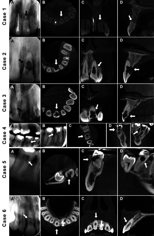
Background: A precise diagnosis is essential for formulating an effective treatment plan for both internal and external resorptions. Cone-beam computed tomography (CBCT) offers a three-dimensional view of the maxillofacial area, capturing images from coronal, axial, and sagittal angles. This method overcomes the limitations of conventional intraoral radiography (IR), especially when it comes to detecting and identifying defects related to internal and external resorption. Thus, the aim of this study was to assess whether CBCT imaging affects the accuracy of diagnosing and planning treatment for internal and external resorption defects differently between endodontic residents (ERs) and specialists (ESs).
Material and methods: Thirty-five clinicians reviewed 3 internal and 3 external resorption cases using clinical histories and intraoral radiographs (IRs), then answered questions about their diagnosis and treatment decisions. One month later, they re-evaluated the cases with CBCT and answered similar questions. Data analyzed using Mc-Nemar chi-square test and Prevalence Adjusted Bias Adjusted Kappa statistic. The level of statistical significance was set off p< 0.05 in all data.
Results: CBCT significantly improved diagnostic accuracy in 2 out of 6 cases (p< 0.001) and altered the treatment plan in 4 out of 6 cases (p< 0.05). There was no significant difference between ERs and ESs regarding diagnosis and treatment planning using the same imaging technique (p> 0.05).
Conclusions: This study suggests that CBCT provides more detailed information compared to IR, with both imaging techniques allowing ERs and ESs to achieve similar diagnostic and treatment planning accuracy. Key words:Clinical decision making, cone-beam computed tomography, dental radiography, diagnosis, root resorption.
期刊介绍:
Indexed in PUBMED, PubMed Central® (PMC) since 2012 and SCOPUSJournal of Clinical and Experimental Dentistry is an Open Access (free access on-line) - http://www.medicinaoral.com/odo/indice.htm. The aim of the Journal of Clinical and Experimental Dentistry is: - Periodontology - Community and Preventive Dentistry - Esthetic Dentistry - Biomaterials and Bioengineering in Dentistry - Operative Dentistry and Endodontics - Prosthetic Dentistry - Orthodontics - Oral Medicine and Pathology - Odontostomatology for the disabled or special patients - Oral Surgery

 求助内容:
求助内容: 应助结果提醒方式:


