Chanhyung Kim, Jisu Son, Dinesh Chaudhary, Yeon-Kyun Park, Ji Hyeon Cho, Dongryeol Ryu, Jee-Heon Jeong, Jonghee Youn
{"title":"通过可视化分析结合可教机器评估干细胞活力。","authors":"Chanhyung Kim, Jisu Son, Dinesh Chaudhary, Yeon-Kyun Park, Ji Hyeon Cho, Dongryeol Ryu, Jee-Heon Jeong, Jonghee Youn","doi":"10.15283/ijsc24105","DOIUrl":null,"url":null,"abstract":"<p><p>Cell viability is an indispensable aspect of cells in the field of drug discovery, cell biology, and biomedical research to assess the physiological conditions of cells such as healthiness, functionality, survivability, etc. Recently, there have been several methods for determining the cell viability through either cell staining with trypan blue and acridine orange, propidium iodide, calcein-AM, etc., or colorimetric assays such as cell counting kit-8 assay. However, these methods have some limitations like time-consuming, expensive, unstable, individual variability, etc. Even present artificial intelligence software such as QuPath, ImageJ, etc., can only determine the cell viability after cell staining. Therefore, we attempted to determine whether cells are alive or not depending on the visual characteristics of an individual cell using Teachable Machine, a web-based artificial intelligence tool provided by Google. Labeling work to assign correct answers to learning data consumes a lot of time and human costs because it is usually done manually. To solve this problem, labeling was automated by recognizing and extracting only individual cells from the image using the contour function to increase time efficiency. In addition, many datasets were created to evaluate and compare the performances of models. Based on the results, the model that showed the best performance showed an accuracy of more than 80%. In conclusion, this model could minimize analysis time, expenses, individual variability, etc., enhancing the efficacy and reproducibility of biological experiments in the fields of drug discovery, drug development, and biological research.</p>","PeriodicalId":14392,"journal":{"name":"International journal of stem cells","volume":" ","pages":"311-319"},"PeriodicalIF":2.4000,"publicationDate":"2025-08-30","publicationTypes":"Journal Article","fieldsOfStudy":null,"isOpenAccess":false,"openAccessPdf":"https://www.ncbi.nlm.nih.gov/pmc/articles/PMC12394080/pdf/","citationCount":"0","resultStr":"{\"title\":\"Assessment of Stem Cell Viability through Visual Analysis Coupled with Teachable Machine.\",\"authors\":\"Chanhyung Kim, Jisu Son, Dinesh Chaudhary, Yeon-Kyun Park, Ji Hyeon Cho, Dongryeol Ryu, Jee-Heon Jeong, Jonghee Youn\",\"doi\":\"10.15283/ijsc24105\",\"DOIUrl\":null,\"url\":null,\"abstract\":\"<p><p>Cell viability is an indispensable aspect of cells in the field of drug discovery, cell biology, and biomedical research to assess the physiological conditions of cells such as healthiness, functionality, survivability, etc. Recently, there have been several methods for determining the cell viability through either cell staining with trypan blue and acridine orange, propidium iodide, calcein-AM, etc., or colorimetric assays such as cell counting kit-8 assay. However, these methods have some limitations like time-consuming, expensive, unstable, individual variability, etc. Even present artificial intelligence software such as QuPath, ImageJ, etc., can only determine the cell viability after cell staining. Therefore, we attempted to determine whether cells are alive or not depending on the visual characteristics of an individual cell using Teachable Machine, a web-based artificial intelligence tool provided by Google. Labeling work to assign correct answers to learning data consumes a lot of time and human costs because it is usually done manually. To solve this problem, labeling was automated by recognizing and extracting only individual cells from the image using the contour function to increase time efficiency. In addition, many datasets were created to evaluate and compare the performances of models. Based on the results, the model that showed the best performance showed an accuracy of more than 80%. In conclusion, this model could minimize analysis time, expenses, individual variability, etc., enhancing the efficacy and reproducibility of biological experiments in the fields of drug discovery, drug development, and biological research.</p>\",\"PeriodicalId\":14392,\"journal\":{\"name\":\"International journal of stem cells\",\"volume\":\" \",\"pages\":\"311-319\"},\"PeriodicalIF\":2.4000,\"publicationDate\":\"2025-08-30\",\"publicationTypes\":\"Journal Article\",\"fieldsOfStudy\":null,\"isOpenAccess\":false,\"openAccessPdf\":\"https://www.ncbi.nlm.nih.gov/pmc/articles/PMC12394080/pdf/\",\"citationCount\":\"0\",\"resultStr\":null,\"platform\":\"Semanticscholar\",\"paperid\":null,\"PeriodicalName\":\"International journal of stem cells\",\"FirstCategoryId\":\"3\",\"ListUrlMain\":\"https://doi.org/10.15283/ijsc24105\",\"RegionNum\":4,\"RegionCategory\":\"医学\",\"ArticlePicture\":[],\"TitleCN\":null,\"AbstractTextCN\":null,\"PMCID\":null,\"EPubDate\":\"2025/6/9 0:00:00\",\"PubModel\":\"Epub\",\"JCR\":\"Q3\",\"JCRName\":\"CELL & TISSUE ENGINEERING\",\"Score\":null,\"Total\":0}","platform":"Semanticscholar","paperid":null,"PeriodicalName":"International journal of stem cells","FirstCategoryId":"3","ListUrlMain":"https://doi.org/10.15283/ijsc24105","RegionNum":4,"RegionCategory":"医学","ArticlePicture":[],"TitleCN":null,"AbstractTextCN":null,"PMCID":null,"EPubDate":"2025/6/9 0:00:00","PubModel":"Epub","JCR":"Q3","JCRName":"CELL & TISSUE ENGINEERING","Score":null,"Total":0}
Assessment of Stem Cell Viability through Visual Analysis Coupled with Teachable Machine.
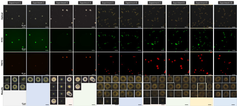
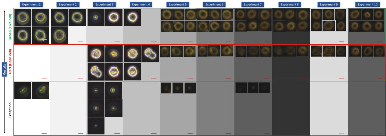
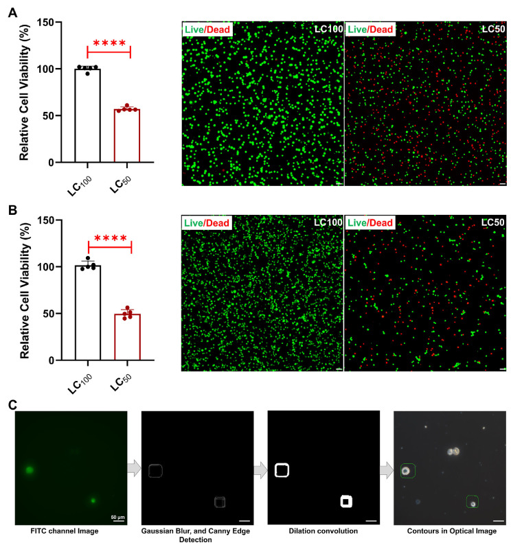
Cell viability is an indispensable aspect of cells in the field of drug discovery, cell biology, and biomedical research to assess the physiological conditions of cells such as healthiness, functionality, survivability, etc. Recently, there have been several methods for determining the cell viability through either cell staining with trypan blue and acridine orange, propidium iodide, calcein-AM, etc., or colorimetric assays such as cell counting kit-8 assay. However, these methods have some limitations like time-consuming, expensive, unstable, individual variability, etc. Even present artificial intelligence software such as QuPath, ImageJ, etc., can only determine the cell viability after cell staining. Therefore, we attempted to determine whether cells are alive or not depending on the visual characteristics of an individual cell using Teachable Machine, a web-based artificial intelligence tool provided by Google. Labeling work to assign correct answers to learning data consumes a lot of time and human costs because it is usually done manually. To solve this problem, labeling was automated by recognizing and extracting only individual cells from the image using the contour function to increase time efficiency. In addition, many datasets were created to evaluate and compare the performances of models. Based on the results, the model that showed the best performance showed an accuracy of more than 80%. In conclusion, this model could minimize analysis time, expenses, individual variability, etc., enhancing the efficacy and reproducibility of biological experiments in the fields of drug discovery, drug development, and biological research.
期刊介绍:
International Journal of Stem Cells (Int J Stem Cells), a peer-reviewed open access journal, principally aims to provide a forum for investigators in the field of stem cell biology to present their research findings and share their visions and opinions. Int J Stem Cells covers all aspects of stem cell biology including basic, clinical and translational research on genetics, biochemistry, and physiology of various types of stem cells including embryonic, adult and induced stem cells. Reports on epigenetics, genomics, proteomics, metabolomics of stem cells are welcome as well. Int J Stem Cells also publishes review articles, technical reports and treatise on ethical issues.
 求助内容:
求助内容: 应助结果提醒方式:


