Davide Allegrini, Raffaele Raimondi, Giovanni Montesano, Marco Caruso, Giovanna Lionetti, Adriano Carnevali, Tania Sorrentino, Vincenzo Scorcia, Mario R Romano
{"title":"特发性视网膜前膜牵引的定量分析:松弛指数的更新版本。","authors":"Davide Allegrini, Raffaele Raimondi, Giovanni Montesano, Marco Caruso, Giovanna Lionetti, Adriano Carnevali, Tania Sorrentino, Vincenzo Scorcia, Mario R Romano","doi":"10.3389/fopht.2025.1528766","DOIUrl":null,"url":null,"abstract":"<p><strong>Purpose: </strong>The aim of this work was to track tangential traction of idiopathic epiretinal membrane from an initial assessment to the immediate post-operative phase using an enhanced version of the relaxation index (RI).</p><p><strong>Methods: </strong>A retrospective analysis was conducted on 9 patients who underwent peeling surgery for idiopathic, symptomatic, and progressive epiretinal membrane. The RI assesses the displacement of vascular crossings in time from a fixed point, which is the retinal pigmented epithelium. This updated iteration integrates infrared images paired with OCT scans instead of OCTA.</p><p><strong>Results: </strong>The study encompassed three timepoints: T1 (initial appointment), T2 (1 week pre-surgery), and Post (1 month post-surgery). T1 was 12± 9 months prior to surgery. A statistically significant difference (p<0.001) in RI was observed across all three timepoints; however, there was no significant correlation between RI and visual acuity (p>0.05).</p><p><strong>Conclusion: </strong>The RI emerges as a comprehensive and direct parameter for objectively assessing and monitoring tangential traction in three dimensions across an extensive area of the posterior pole. Further streamlining of the process is necessary to integrate this feature into clinical practice effectively.</p>","PeriodicalId":73096,"journal":{"name":"Frontiers in ophthalmology","volume":"5 ","pages":"1528766"},"PeriodicalIF":0.9000,"publicationDate":"2025-05-22","publicationTypes":"Journal Article","fieldsOfStudy":null,"isOpenAccess":false,"openAccessPdf":"https://www.ncbi.nlm.nih.gov/pmc/articles/PMC12137094/pdf/","citationCount":"0","resultStr":"{\"title\":\"Quantitative analysis of idiopathic epiretinal membrane traction: an updated version of the relaxation index.\",\"authors\":\"Davide Allegrini, Raffaele Raimondi, Giovanni Montesano, Marco Caruso, Giovanna Lionetti, Adriano Carnevali, Tania Sorrentino, Vincenzo Scorcia, Mario R Romano\",\"doi\":\"10.3389/fopht.2025.1528766\",\"DOIUrl\":null,\"url\":null,\"abstract\":\"<p><strong>Purpose: </strong>The aim of this work was to track tangential traction of idiopathic epiretinal membrane from an initial assessment to the immediate post-operative phase using an enhanced version of the relaxation index (RI).</p><p><strong>Methods: </strong>A retrospective analysis was conducted on 9 patients who underwent peeling surgery for idiopathic, symptomatic, and progressive epiretinal membrane. The RI assesses the displacement of vascular crossings in time from a fixed point, which is the retinal pigmented epithelium. This updated iteration integrates infrared images paired with OCT scans instead of OCTA.</p><p><strong>Results: </strong>The study encompassed three timepoints: T1 (initial appointment), T2 (1 week pre-surgery), and Post (1 month post-surgery). T1 was 12± 9 months prior to surgery. A statistically significant difference (p<0.001) in RI was observed across all three timepoints; however, there was no significant correlation between RI and visual acuity (p>0.05).</p><p><strong>Conclusion: </strong>The RI emerges as a comprehensive and direct parameter for objectively assessing and monitoring tangential traction in three dimensions across an extensive area of the posterior pole. Further streamlining of the process is necessary to integrate this feature into clinical practice effectively.</p>\",\"PeriodicalId\":73096,\"journal\":{\"name\":\"Frontiers in ophthalmology\",\"volume\":\"5 \",\"pages\":\"1528766\"},\"PeriodicalIF\":0.9000,\"publicationDate\":\"2025-05-22\",\"publicationTypes\":\"Journal Article\",\"fieldsOfStudy\":null,\"isOpenAccess\":false,\"openAccessPdf\":\"https://www.ncbi.nlm.nih.gov/pmc/articles/PMC12137094/pdf/\",\"citationCount\":\"0\",\"resultStr\":null,\"platform\":\"Semanticscholar\",\"paperid\":null,\"PeriodicalName\":\"Frontiers in ophthalmology\",\"FirstCategoryId\":\"1085\",\"ListUrlMain\":\"https://doi.org/10.3389/fopht.2025.1528766\",\"RegionNum\":0,\"RegionCategory\":null,\"ArticlePicture\":[],\"TitleCN\":null,\"AbstractTextCN\":null,\"PMCID\":null,\"EPubDate\":\"2025/1/1 0:00:00\",\"PubModel\":\"eCollection\",\"JCR\":\"\",\"JCRName\":\"\",\"Score\":null,\"Total\":0}","platform":"Semanticscholar","paperid":null,"PeriodicalName":"Frontiers in ophthalmology","FirstCategoryId":"1085","ListUrlMain":"https://doi.org/10.3389/fopht.2025.1528766","RegionNum":0,"RegionCategory":null,"ArticlePicture":[],"TitleCN":null,"AbstractTextCN":null,"PMCID":null,"EPubDate":"2025/1/1 0:00:00","PubModel":"eCollection","JCR":"","JCRName":"","Score":null,"Total":0}
Quantitative analysis of idiopathic epiretinal membrane traction: an updated version of the relaxation index.
Purpose: The aim of this work was to track tangential traction of idiopathic epiretinal membrane from an initial assessment to the immediate post-operative phase using an enhanced version of the relaxation index (RI).
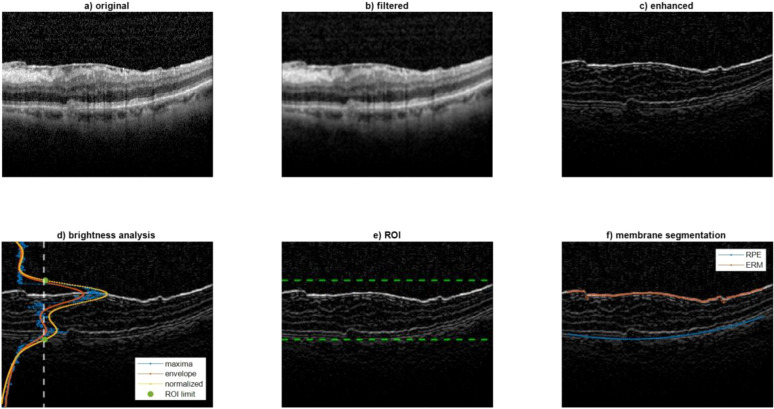
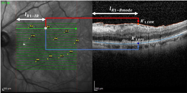
Methods: A retrospective analysis was conducted on 9 patients who underwent peeling surgery for idiopathic, symptomatic, and progressive epiretinal membrane. The RI assesses the displacement of vascular crossings in time from a fixed point, which is the retinal pigmented epithelium. This updated iteration integrates infrared images paired with OCT scans instead of OCTA.
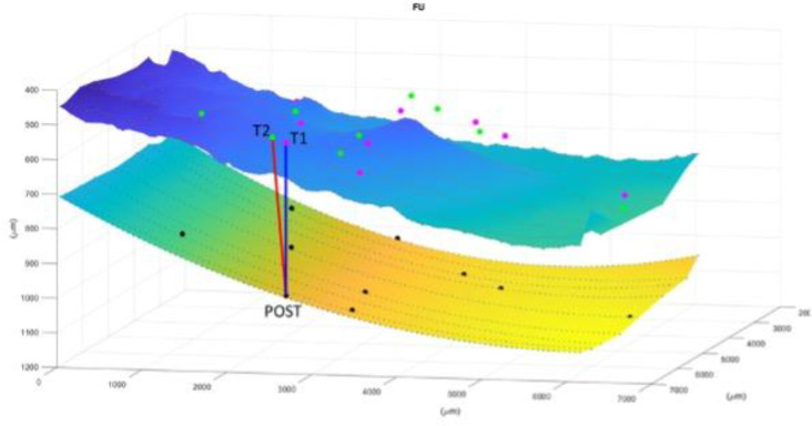
Results: The study encompassed three timepoints: T1 (initial appointment), T2 (1 week pre-surgery), and Post (1 month post-surgery). T1 was 12± 9 months prior to surgery. A statistically significant difference (p<0.001) in RI was observed across all three timepoints; however, there was no significant correlation between RI and visual acuity (p>0.05).
Conclusion: The RI emerges as a comprehensive and direct parameter for objectively assessing and monitoring tangential traction in three dimensions across an extensive area of the posterior pole. Further streamlining of the process is necessary to integrate this feature into clinical practice effectively.

 求助内容:
求助内容: 应助结果提醒方式:


