{"title":"痛风成像:地图集。","authors":"Luqman Wali, Emma Rowbotham","doi":"10.1093/rap/rkaf051","DOIUrl":null,"url":null,"abstract":"<p><p>Gout is a common systemic disease defined by deposition of monosodium urate (MSU) crystals in articular and peri-articular structures, leading to recurrent bouts of inflammation. Imaging plays an important role in establishing the diagnosis when crystal aspiration is not feasible and the clinical diagnosis is uncertain. Each imaging modality has a unique role. Radiographs can demonstrate characteristic erosions and tophi in later stages of gout. Ultrasound has a major role in the diagnosis and assessment of gout. Dual-energy computed tomography (DECT) enables precise visualization of MSU deposits and can determine disease burden. MRI can assess for non-specific inflammatory and structural changes. Both ultrasound and DECT are emphasized as part of diagnostic algorithms and the role of imaging is expanding with more recent advancements and evidence. This review provides an imaging-centric overview of each modality and its evolving significance in gout.</p>","PeriodicalId":21350,"journal":{"name":"Rheumatology Advances in Practice","volume":"9 2","pages":"rkaf051"},"PeriodicalIF":2.1000,"publicationDate":"2025-06-04","publicationTypes":"Journal Article","fieldsOfStudy":null,"isOpenAccess":false,"openAccessPdf":"https://www.ncbi.nlm.nih.gov/pmc/articles/PMC12137905/pdf/","citationCount":"0","resultStr":"{\"title\":\"Imaging of gout: an atlas.\",\"authors\":\"Luqman Wali, Emma Rowbotham\",\"doi\":\"10.1093/rap/rkaf051\",\"DOIUrl\":null,\"url\":null,\"abstract\":\"<p><p>Gout is a common systemic disease defined by deposition of monosodium urate (MSU) crystals in articular and peri-articular structures, leading to recurrent bouts of inflammation. Imaging plays an important role in establishing the diagnosis when crystal aspiration is not feasible and the clinical diagnosis is uncertain. Each imaging modality has a unique role. Radiographs can demonstrate characteristic erosions and tophi in later stages of gout. Ultrasound has a major role in the diagnosis and assessment of gout. Dual-energy computed tomography (DECT) enables precise visualization of MSU deposits and can determine disease burden. MRI can assess for non-specific inflammatory and structural changes. Both ultrasound and DECT are emphasized as part of diagnostic algorithms and the role of imaging is expanding with more recent advancements and evidence. This review provides an imaging-centric overview of each modality and its evolving significance in gout.</p>\",\"PeriodicalId\":21350,\"journal\":{\"name\":\"Rheumatology Advances in Practice\",\"volume\":\"9 2\",\"pages\":\"rkaf051\"},\"PeriodicalIF\":2.1000,\"publicationDate\":\"2025-06-04\",\"publicationTypes\":\"Journal Article\",\"fieldsOfStudy\":null,\"isOpenAccess\":false,\"openAccessPdf\":\"https://www.ncbi.nlm.nih.gov/pmc/articles/PMC12137905/pdf/\",\"citationCount\":\"0\",\"resultStr\":null,\"platform\":\"Semanticscholar\",\"paperid\":null,\"PeriodicalName\":\"Rheumatology Advances in Practice\",\"FirstCategoryId\":\"1085\",\"ListUrlMain\":\"https://doi.org/10.1093/rap/rkaf051\",\"RegionNum\":0,\"RegionCategory\":null,\"ArticlePicture\":[],\"TitleCN\":null,\"AbstractTextCN\":null,\"PMCID\":null,\"EPubDate\":\"2025/1/1 0:00:00\",\"PubModel\":\"eCollection\",\"JCR\":\"Q3\",\"JCRName\":\"RHEUMATOLOGY\",\"Score\":null,\"Total\":0}","platform":"Semanticscholar","paperid":null,"PeriodicalName":"Rheumatology Advances in Practice","FirstCategoryId":"1085","ListUrlMain":"https://doi.org/10.1093/rap/rkaf051","RegionNum":0,"RegionCategory":null,"ArticlePicture":[],"TitleCN":null,"AbstractTextCN":null,"PMCID":null,"EPubDate":"2025/1/1 0:00:00","PubModel":"eCollection","JCR":"Q3","JCRName":"RHEUMATOLOGY","Score":null,"Total":0}
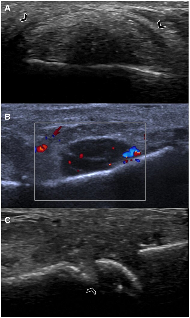
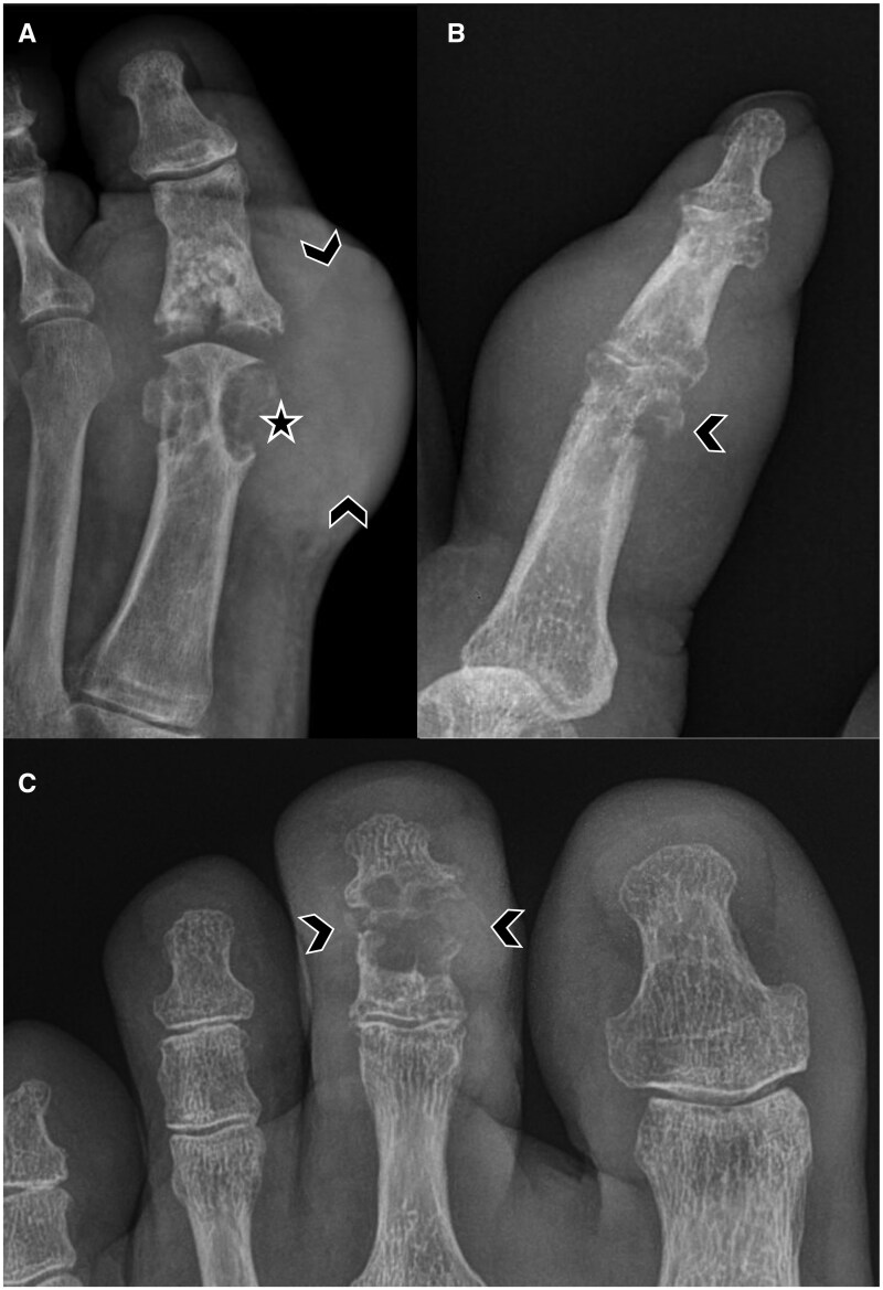
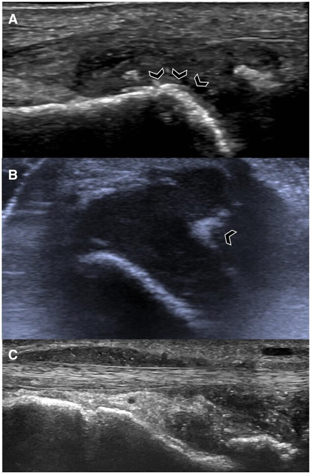
Gout is a common systemic disease defined by deposition of monosodium urate (MSU) crystals in articular and peri-articular structures, leading to recurrent bouts of inflammation. Imaging plays an important role in establishing the diagnosis when crystal aspiration is not feasible and the clinical diagnosis is uncertain. Each imaging modality has a unique role. Radiographs can demonstrate characteristic erosions and tophi in later stages of gout. Ultrasound has a major role in the diagnosis and assessment of gout. Dual-energy computed tomography (DECT) enables precise visualization of MSU deposits and can determine disease burden. MRI can assess for non-specific inflammatory and structural changes. Both ultrasound and DECT are emphasized as part of diagnostic algorithms and the role of imaging is expanding with more recent advancements and evidence. This review provides an imaging-centric overview of each modality and its evolving significance in gout.

 求助内容:
求助内容: 应助结果提醒方式:


