Mohd N Mat Nor, Colin R Green, David Squirrell, Monica L Acosta
{"title":"视网膜高反射灶是眼部疾病的生物标志物:基于人类证据和动物模型的综述","authors":"Mohd N Mat Nor, Colin R Green, David Squirrell, Monica L Acosta","doi":"10.1155/joph/9573587","DOIUrl":null,"url":null,"abstract":"<p><p><b>Background:</b> Abnormalities in the retina have a profound impact on vision, and accurate diagnosis and monitoring are essential for effective clinical management. Retinal hyperreflective foci (HRF), lesions, or dots, identified using optical coherence tomography (OCT), are observed in both animals and humans and have been associated with several ocular conditions, including diabetic retinopathy (DR), age-related macular degeneration (AMD), and retinal vascular diseases. <b>Methods:</b> To evaluate the relevance of retinal HRF, we conducted a comprehensive scoping review of the literature published up to July 2024 including in the discussion key papers that emerged in 2025. Our search spanned electronic databases utilizing carefully identified search terms related to HRF and OCT within the last six years. We excluded publications on HRF outside the retina, treatments, non-peer-reviewed content, duplicates, studies older than 6 years, and those not focused on AMD, DR, or glaucoma. <b>Results:</b> A total of 141,085 records were initially identified from various databases and further refined based on keywords and content relevance. Finally, 42 reports meeting the criteria were retained for in-depth analysis. HRF were observed mainly in OCT scans of the AMD retina, as well as in DR and, to a lesser extent, in other retinopathies and interestingly in glaucoma. In AMD, HRF are described as a marker for disease progression, often associated with a compromised photoreceptor structure. In DR, HRF indicated issues such as abnormal blood vessels and cellular changes linked to microglia activation. In glaucoma, HRF may reflect microglia and macrophage activation. Most publications concur that the presence of HRF correlates with inflammatory processes and aging in the retina, with early appearance of small HRF serving as a biomarker for ocular disease. The size of HRF and their location were consistent with disease presentation. <b>Conclusion:</b> There is an agreement that HRF of less than 30 μm are biomarkers of inflammation in the retina despite having variable intraretinal locations. HRF resulting from the effect of aging can be discerned from AMD based on their quantity and appearance. The results show the importance of HRF as a biomarker of ocular disease and confirm that HRF are indicative of an inflammatory eye disorder.</p>","PeriodicalId":16674,"journal":{"name":"Journal of Ophthalmology","volume":"2025 ","pages":"9573587"},"PeriodicalIF":1.9000,"publicationDate":"2025-05-29","publicationTypes":"Journal Article","fieldsOfStudy":null,"isOpenAccess":false,"openAccessPdf":"https://www.ncbi.nlm.nih.gov/pmc/articles/PMC12140829/pdf/","citationCount":"0","resultStr":"{\"title\":\"Retinal Hyperreflective Foci Are Biomarkers of Ocular Disease: A Scoping Review With Evidence From Humans and Insights From Animal Models.\",\"authors\":\"Mohd N Mat Nor, Colin R Green, David Squirrell, Monica L Acosta\",\"doi\":\"10.1155/joph/9573587\",\"DOIUrl\":null,\"url\":null,\"abstract\":\"<p><p><b>Background:</b> Abnormalities in the retina have a profound impact on vision, and accurate diagnosis and monitoring are essential for effective clinical management. Retinal hyperreflective foci (HRF), lesions, or dots, identified using optical coherence tomography (OCT), are observed in both animals and humans and have been associated with several ocular conditions, including diabetic retinopathy (DR), age-related macular degeneration (AMD), and retinal vascular diseases. <b>Methods:</b> To evaluate the relevance of retinal HRF, we conducted a comprehensive scoping review of the literature published up to July 2024 including in the discussion key papers that emerged in 2025. Our search spanned electronic databases utilizing carefully identified search terms related to HRF and OCT within the last six years. We excluded publications on HRF outside the retina, treatments, non-peer-reviewed content, duplicates, studies older than 6 years, and those not focused on AMD, DR, or glaucoma. <b>Results:</b> A total of 141,085 records were initially identified from various databases and further refined based on keywords and content relevance. Finally, 42 reports meeting the criteria were retained for in-depth analysis. HRF were observed mainly in OCT scans of the AMD retina, as well as in DR and, to a lesser extent, in other retinopathies and interestingly in glaucoma. In AMD, HRF are described as a marker for disease progression, often associated with a compromised photoreceptor structure. In DR, HRF indicated issues such as abnormal blood vessels and cellular changes linked to microglia activation. In glaucoma, HRF may reflect microglia and macrophage activation. Most publications concur that the presence of HRF correlates with inflammatory processes and aging in the retina, with early appearance of small HRF serving as a biomarker for ocular disease. The size of HRF and their location were consistent with disease presentation. <b>Conclusion:</b> There is an agreement that HRF of less than 30 μm are biomarkers of inflammation in the retina despite having variable intraretinal locations. HRF resulting from the effect of aging can be discerned from AMD based on their quantity and appearance. The results show the importance of HRF as a biomarker of ocular disease and confirm that HRF are indicative of an inflammatory eye disorder.</p>\",\"PeriodicalId\":16674,\"journal\":{\"name\":\"Journal of Ophthalmology\",\"volume\":\"2025 \",\"pages\":\"9573587\"},\"PeriodicalIF\":1.9000,\"publicationDate\":\"2025-05-29\",\"publicationTypes\":\"Journal Article\",\"fieldsOfStudy\":null,\"isOpenAccess\":false,\"openAccessPdf\":\"https://www.ncbi.nlm.nih.gov/pmc/articles/PMC12140829/pdf/\",\"citationCount\":\"0\",\"resultStr\":null,\"platform\":\"Semanticscholar\",\"paperid\":null,\"PeriodicalName\":\"Journal of Ophthalmology\",\"FirstCategoryId\":\"3\",\"ListUrlMain\":\"https://doi.org/10.1155/joph/9573587\",\"RegionNum\":4,\"RegionCategory\":\"医学\",\"ArticlePicture\":[],\"TitleCN\":null,\"AbstractTextCN\":null,\"PMCID\":null,\"EPubDate\":\"2025/1/1 0:00:00\",\"PubModel\":\"eCollection\",\"JCR\":\"Q3\",\"JCRName\":\"OPHTHALMOLOGY\",\"Score\":null,\"Total\":0}","platform":"Semanticscholar","paperid":null,"PeriodicalName":"Journal of Ophthalmology","FirstCategoryId":"3","ListUrlMain":"https://doi.org/10.1155/joph/9573587","RegionNum":4,"RegionCategory":"医学","ArticlePicture":[],"TitleCN":null,"AbstractTextCN":null,"PMCID":null,"EPubDate":"2025/1/1 0:00:00","PubModel":"eCollection","JCR":"Q3","JCRName":"OPHTHALMOLOGY","Score":null,"Total":0}
Retinal Hyperreflective Foci Are Biomarkers of Ocular Disease: A Scoping Review With Evidence From Humans and Insights From Animal Models.
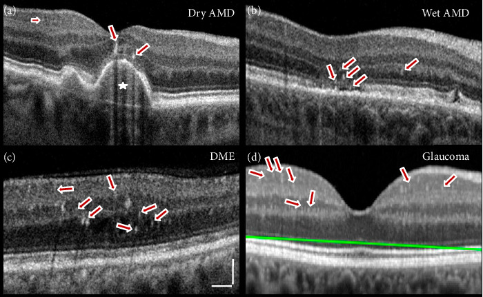
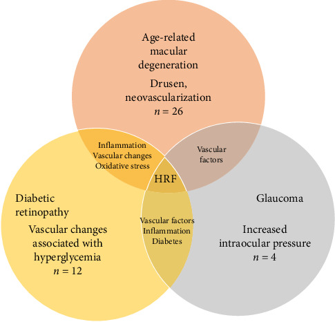
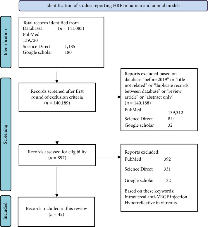
Background: Abnormalities in the retina have a profound impact on vision, and accurate diagnosis and monitoring are essential for effective clinical management. Retinal hyperreflective foci (HRF), lesions, or dots, identified using optical coherence tomography (OCT), are observed in both animals and humans and have been associated with several ocular conditions, including diabetic retinopathy (DR), age-related macular degeneration (AMD), and retinal vascular diseases. Methods: To evaluate the relevance of retinal HRF, we conducted a comprehensive scoping review of the literature published up to July 2024 including in the discussion key papers that emerged in 2025. Our search spanned electronic databases utilizing carefully identified search terms related to HRF and OCT within the last six years. We excluded publications on HRF outside the retina, treatments, non-peer-reviewed content, duplicates, studies older than 6 years, and those not focused on AMD, DR, or glaucoma. Results: A total of 141,085 records were initially identified from various databases and further refined based on keywords and content relevance. Finally, 42 reports meeting the criteria were retained for in-depth analysis. HRF were observed mainly in OCT scans of the AMD retina, as well as in DR and, to a lesser extent, in other retinopathies and interestingly in glaucoma. In AMD, HRF are described as a marker for disease progression, often associated with a compromised photoreceptor structure. In DR, HRF indicated issues such as abnormal blood vessels and cellular changes linked to microglia activation. In glaucoma, HRF may reflect microglia and macrophage activation. Most publications concur that the presence of HRF correlates with inflammatory processes and aging in the retina, with early appearance of small HRF serving as a biomarker for ocular disease. The size of HRF and their location were consistent with disease presentation. Conclusion: There is an agreement that HRF of less than 30 μm are biomarkers of inflammation in the retina despite having variable intraretinal locations. HRF resulting from the effect of aging can be discerned from AMD based on their quantity and appearance. The results show the importance of HRF as a biomarker of ocular disease and confirm that HRF are indicative of an inflammatory eye disorder.
期刊介绍:
Journal of Ophthalmology is a peer-reviewed, Open Access journal that publishes original research articles, review articles, and clinical studies related to the anatomy, physiology and diseases of the eye. Submissions should focus on new diagnostic and surgical techniques, instrument and therapy updates, as well as clinical trials and research findings.
 求助内容:
求助内容: 应助结果提醒方式:


