{"title":"脂肪肉瘤27例临床病理及分子诊断特点分析。","authors":"Yin Zhu, Dong Chen, Jingjing Yu, Shuo Wang","doi":"10.25259/Cytojournal_246_2024","DOIUrl":null,"url":null,"abstract":"<p><strong>Objective: </strong>Liposarcomas are rare tumors, and it is difficult to collect cases in less densely populated areas. Therefore, we aimed to document more cases over a relatively long period to provide more data about the characteristics of liposarcomas. In this study, the clinicopathological features of liposarcomas were investigated in 27 patients.</p><p><strong>Material and methods: </strong>All cases were confirmed by diagnosis through hematoxylin and eosin staining, immunohistochemistry (IHC), and fluorescence <i>in situ</i> hybridization (FISH). Combined IHC analysis was performed for murine double minute 2 (MDM2), cyclin-dependent kinase 4 (CDK4), multiple tumor suppressor 1 (P16), and Cyclin D1. FISH was performed to detect MDM2 amplification in atypical lipomatous tumor/well-differentiated liposarcoma (ALT/WDLPS) and dedifferentiated liposarcoma (DDLPS), and DNA damage inducible transcript 3 ( DDIT3) rearrangements in myxoid liposarcoma (MLPS).</p><p><strong>Results: </strong>Seven cases of liposarcoma were located in the paratesticular region (25.9%, 7/27), 12 in the retroperitoneum (44.4%, 12/27), and eight in the limbs (29.6%, 8/27). Histological analysis showed that there were 13 cases of ALT/WDLPS (48.1%, 13/27), nine cases of DDLPS (33.3%, 9/27), three cases of MLPS (11.1%, 3/27), and two cases of pleomorphic liposarcoma (7.4%, 2/27). IHC analysis revealed that 26 cases were MDM2-positive (96.3%, 26/27), 22 were CDK4-positive (81.5%, 22/27), 26 were P16-positive (96.3%, 26/27), and 27 were cyclin D1-positive (100%, 27/27). FISH analysis revealed 20 cases of MDM2 positivity (90.9%, 20/22) and one case of DDIT3 positivity (50%, 1/2). The clinical outcomes were available for 21 patients. Four patients died (4/21, 19.0%), five experienced recurrence (5/21, 23.8%), and 12 (12/21, 57.1%) survived with no other disease.</p><p><strong>Conclusion: </strong>A combined IHC examination of the four indicators may be used to diagnose ALT/WDLPS and DDLPS, and FISH is recommended as an important supporting method.</p>","PeriodicalId":49082,"journal":{"name":"Cytojournal","volume":"22 ","pages":"40"},"PeriodicalIF":3.1000,"publicationDate":"2025-04-01","publicationTypes":"Journal Article","fieldsOfStudy":null,"isOpenAccess":false,"openAccessPdf":"https://www.ncbi.nlm.nih.gov/pmc/articles/PMC12134912/pdf/","citationCount":"0","resultStr":"{\"title\":\"Clinicopathological and molecular diagnostic features of liposarcoma: A study of 27 cases.\",\"authors\":\"Yin Zhu, Dong Chen, Jingjing Yu, Shuo Wang\",\"doi\":\"10.25259/Cytojournal_246_2024\",\"DOIUrl\":null,\"url\":null,\"abstract\":\"<p><strong>Objective: </strong>Liposarcomas are rare tumors, and it is difficult to collect cases in less densely populated areas. Therefore, we aimed to document more cases over a relatively long period to provide more data about the characteristics of liposarcomas. In this study, the clinicopathological features of liposarcomas were investigated in 27 patients.</p><p><strong>Material and methods: </strong>All cases were confirmed by diagnosis through hematoxylin and eosin staining, immunohistochemistry (IHC), and fluorescence <i>in situ</i> hybridization (FISH). Combined IHC analysis was performed for murine double minute 2 (MDM2), cyclin-dependent kinase 4 (CDK4), multiple tumor suppressor 1 (P16), and Cyclin D1. FISH was performed to detect MDM2 amplification in atypical lipomatous tumor/well-differentiated liposarcoma (ALT/WDLPS) and dedifferentiated liposarcoma (DDLPS), and DNA damage inducible transcript 3 ( DDIT3) rearrangements in myxoid liposarcoma (MLPS).</p><p><strong>Results: </strong>Seven cases of liposarcoma were located in the paratesticular region (25.9%, 7/27), 12 in the retroperitoneum (44.4%, 12/27), and eight in the limbs (29.6%, 8/27). Histological analysis showed that there were 13 cases of ALT/WDLPS (48.1%, 13/27), nine cases of DDLPS (33.3%, 9/27), three cases of MLPS (11.1%, 3/27), and two cases of pleomorphic liposarcoma (7.4%, 2/27). IHC analysis revealed that 26 cases were MDM2-positive (96.3%, 26/27), 22 were CDK4-positive (81.5%, 22/27), 26 were P16-positive (96.3%, 26/27), and 27 were cyclin D1-positive (100%, 27/27). FISH analysis revealed 20 cases of MDM2 positivity (90.9%, 20/22) and one case of DDIT3 positivity (50%, 1/2). The clinical outcomes were available for 21 patients. Four patients died (4/21, 19.0%), five experienced recurrence (5/21, 23.8%), and 12 (12/21, 57.1%) survived with no other disease.</p><p><strong>Conclusion: </strong>A combined IHC examination of the four indicators may be used to diagnose ALT/WDLPS and DDLPS, and FISH is recommended as an important supporting method.</p>\",\"PeriodicalId\":49082,\"journal\":{\"name\":\"Cytojournal\",\"volume\":\"22 \",\"pages\":\"40\"},\"PeriodicalIF\":3.1000,\"publicationDate\":\"2025-04-01\",\"publicationTypes\":\"Journal Article\",\"fieldsOfStudy\":null,\"isOpenAccess\":false,\"openAccessPdf\":\"https://www.ncbi.nlm.nih.gov/pmc/articles/PMC12134912/pdf/\",\"citationCount\":\"0\",\"resultStr\":null,\"platform\":\"Semanticscholar\",\"paperid\":null,\"PeriodicalName\":\"Cytojournal\",\"FirstCategoryId\":\"3\",\"ListUrlMain\":\"https://doi.org/10.25259/Cytojournal_246_2024\",\"RegionNum\":4,\"RegionCategory\":\"医学\",\"ArticlePicture\":[],\"TitleCN\":null,\"AbstractTextCN\":null,\"PMCID\":null,\"EPubDate\":\"2025/1/1 0:00:00\",\"PubModel\":\"eCollection\",\"JCR\":\"Q2\",\"JCRName\":\"PATHOLOGY\",\"Score\":null,\"Total\":0}","platform":"Semanticscholar","paperid":null,"PeriodicalName":"Cytojournal","FirstCategoryId":"3","ListUrlMain":"https://doi.org/10.25259/Cytojournal_246_2024","RegionNum":4,"RegionCategory":"医学","ArticlePicture":[],"TitleCN":null,"AbstractTextCN":null,"PMCID":null,"EPubDate":"2025/1/1 0:00:00","PubModel":"eCollection","JCR":"Q2","JCRName":"PATHOLOGY","Score":null,"Total":0}
Clinicopathological and molecular diagnostic features of liposarcoma: A study of 27 cases.
Objective: Liposarcomas are rare tumors, and it is difficult to collect cases in less densely populated areas. Therefore, we aimed to document more cases over a relatively long period to provide more data about the characteristics of liposarcomas. In this study, the clinicopathological features of liposarcomas were investigated in 27 patients.
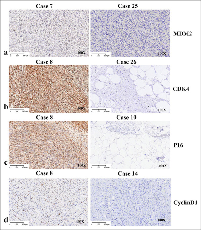
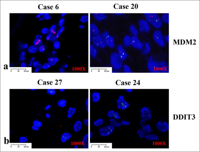
Material and methods: All cases were confirmed by diagnosis through hematoxylin and eosin staining, immunohistochemistry (IHC), and fluorescence in situ hybridization (FISH). Combined IHC analysis was performed for murine double minute 2 (MDM2), cyclin-dependent kinase 4 (CDK4), multiple tumor suppressor 1 (P16), and Cyclin D1. FISH was performed to detect MDM2 amplification in atypical lipomatous tumor/well-differentiated liposarcoma (ALT/WDLPS) and dedifferentiated liposarcoma (DDLPS), and DNA damage inducible transcript 3 ( DDIT3) rearrangements in myxoid liposarcoma (MLPS).
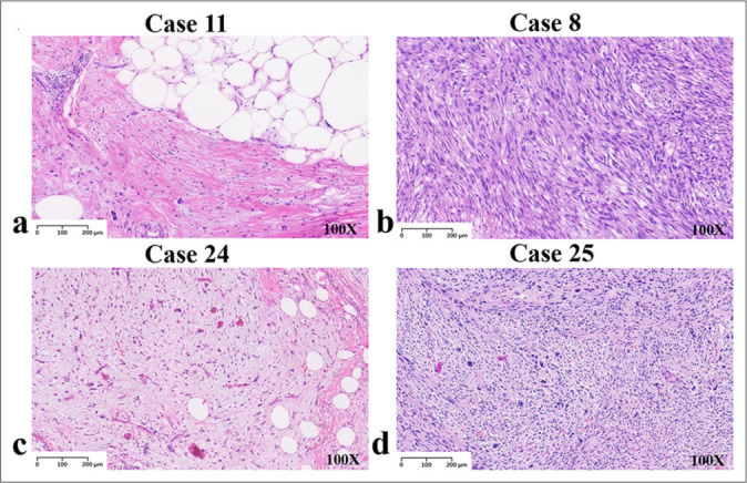
Results: Seven cases of liposarcoma were located in the paratesticular region (25.9%, 7/27), 12 in the retroperitoneum (44.4%, 12/27), and eight in the limbs (29.6%, 8/27). Histological analysis showed that there were 13 cases of ALT/WDLPS (48.1%, 13/27), nine cases of DDLPS (33.3%, 9/27), three cases of MLPS (11.1%, 3/27), and two cases of pleomorphic liposarcoma (7.4%, 2/27). IHC analysis revealed that 26 cases were MDM2-positive (96.3%, 26/27), 22 were CDK4-positive (81.5%, 22/27), 26 were P16-positive (96.3%, 26/27), and 27 were cyclin D1-positive (100%, 27/27). FISH analysis revealed 20 cases of MDM2 positivity (90.9%, 20/22) and one case of DDIT3 positivity (50%, 1/2). The clinical outcomes were available for 21 patients. Four patients died (4/21, 19.0%), five experienced recurrence (5/21, 23.8%), and 12 (12/21, 57.1%) survived with no other disease.
Conclusion: A combined IHC examination of the four indicators may be used to diagnose ALT/WDLPS and DDLPS, and FISH is recommended as an important supporting method.
期刊介绍:
The CytoJournal is an open-access peer-reviewed journal committed to publishing high-quality articles in the field of Diagnostic Cytopathology including Molecular aspects. The journal is owned by the Cytopathology Foundation and published by the Scientific Scholar.
 求助内容:
求助内容: 应助结果提醒方式:


